Download
1 / 31
330 likes | 421 Vues
Lymphadenopathy and Malignancy. Outline. Introducing Historical Clues Physical Examination Nodal Character and Size Diagnosis and Management Lymph Node Biopsy. Introducing. Lymphadenopathy : an abnormality in the size or character of lymph nodes

E N D
Outline • Introducing • Historical Clues • Physical Examination • Nodal Character and Size • Diagnosis and Management • Lymph Node Biopsy
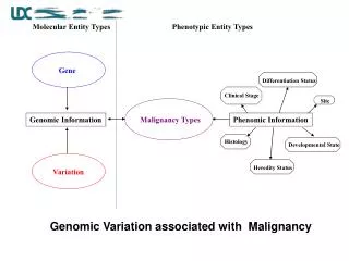
Introducing • Lymphadenopathy : an abnormality in the size or character of lymph nodes • Categories of Lymphadenopathy : MIAMI Malignancies, Infections, Autoimmune disorders, Miscellaneous and unusual conditions, and Iatrogenic causes • The most concerning to the patient and physician : the possibility of underlying malignancy • Low Rate of Malignancy in Primary Care : 1.1 % of pt’s presenting to the office with unexplained lymphadenopathy
Historical Clues • Age and Duration • Exposures & History • Associated Symptoms
Historical Clues : Age and Duration • Malignant rate increases with age. • A majority of healthy children have palpable cervical, inguinal and axillary adenopathy. Most of them is infectious or benign in etiology. • Lymphadenopathy that lasts less than 2 weeks or more than 1 year with no progressive size increase has a very low likelihood of being neoplastic. • Rare Exception : low-grade Hodgkin’s/ non-Hodgkin’s lymphomas and, occasionally, chronic lymphocytic leukemia
Historical Clues : Exposures & History • Animals, biting insects, infectious contacts, recurrent infections, chronic use of medications. Travel-related exposures and immunization status. • Tobacco, alcohol, ultraviolet radiation : raise suspicion for metastatic carcinoma • Occupational exposures to silicon or beryllium • Sexual history and orientation. AIDS pt’s • Family history
Medications That Can Cause Lymphadenopathy • Allopurinol (Zyloprim) • Atenolol (Tenormin) • Captopril (Capoten) • Carbamazepine (Tegretol) • Gold • Hydralazine (Apresoline) • Penicillins • Phenytoin (Dilantin) • Primidone (Mysoline) • Pyrimethamine (Daraprim) • Quinidine • Trimethoprim/sulfamethoxazole (Bactrim) • Sulindac (Clinoril)
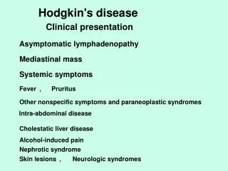
Historical Clues : Associated Symptoms • Constitutional symptoms : fever, fatigue, malaise with atypical lymphocytosis → mononucleosis syndromes • Significant fever, night sweats, unexplained BW loss > 10% of normal BW → “B” symptoms of Hodgkin’s lymphoma • Arthralgias, muscle weakness, unusual rash → autoimmune diseases such as RA, SLE, dermatomyositis
Physical Examination • Head and Neck LN • Axillary LN • Inguinal LN
Lymph nodes of the head and neck, and the regions that they drain
Head and Neck Lymphadenopathy • In one outpatient primary care study : cervical LNs are palpable in 51% of adult physicals, with the incidence declined with age. • Infection is the most common cause • Most cases resolve quickly; some entities can create persistent lymphadenopathy for months. (ex. Atypical mycobacteria, cat-scratch disease, toxoplasmosis, kikuchi’s lymphadenitis, sarcoidosis, Kawasaki’s syndrome.) • Supraclavicular nodes are the most likely to be malignant and should always be investigated, even in children.
Axillary Lymphadenopathy • Most of cases are nonspecific or reactive to local injury/infection in etiology. • Persistent lymphadenopathy is less commonly found in the axillary nodes than in the inguinal chain. • Breast adenocarcinoma often metastasis initially to the anterior and central axillary nodes, which may be palpable before discovery of the primary tumor. • Antecubital or epitrochlear lymphadenopathy can suggest lymphoma or melanoma of the extremity.
Inguinal Lymphadenopathy • It is common, with nodes enlarged up to 1 to 2 cm in diameter in many healthy adults, but it is of low suspicion of malignancy. • Benign reactive lymphadenopathy and infection are the most common etiologies. • Although some tumors, such as Hodgkin’s lymphomas, penile/ vulvar SCC, melanoma in this area, may present with inguinal lymphadenopathy, it is typical presenting finding in neither case.
Generalized Lymphadenopathy • Generalized lymphadenopathy : lymphadenopathy found in two or more distinct anatomic regions • More likely to result from serious infections, autoimmune diseases, and disseminated malignancies. • Specific testing is usually required. • Generalized adenopathy infrequently occurs in pt’s with neoplasms, but it is occasionally seen in patients with leukemias and lymphomas, or advanced disseminated metastatic solid tumors.
Nodal Character and Size • Hard and painless nodes have higher suspicion of malignancy or granulomatous disease. • Viral infection typically produces hyperplastic nodes that are bilateral, mobile, nontender, and clearly demarcated. • Palpable supraclavicular, iliac, or popliteal nodes of any size and epitrochlear nodes larger than 5mm are considered abnormal. • Increasing size and persistence over time are of greater concern for malignancy than a specific level of nodal enlargement.
Diagnosis and Management • The first step : reviewing pts’ medications, considering unusual causes of lymphadenopathy, and reconsidering the risk factors for neoplasm. If a diagnosis is not suggested, and the patient is deemed low risk for neoplasm, the regional lymphadenopathy can be safely observed. • It is suggested that non-inguinal lymphadenopathy lasting more than one month merits specific investigation or biopsy.
Lymph Node Biopsy • Once biopsy has been chosen, ideally the largest, most suspicious, and most accessible node is selected, taking into account differing diagnostic yields by site. • Inguinal nodes offer the lowest yield, and supraclavicular nodes have the highest. • Excisional biopsy remains the diagnostic procedure of choice.
Persistent Generalized Lymphadenopathy (PGL) Presenting Signs and Symptoms • Lymph nodes larger than 1.5 cm in diameter in 2 or more extrainguinal sites of 3 or more months duration • Nodes are non-tender, symmetrical, and often involve the posterior cervical, axillary, occipital, and epitrochlear nodes
Overview • Swelling of lymph nodes is a frequently encountered symptom • It is important to carry out a careful history and physical exam • The cause often becomes obvious, but in more complicated cases, laboratory tests and lymph node biopsy may be necessary to establish a definitive diagnosis
Diagnostics • Where possible, do a CBC (FBC) and chest x-ray before making a diagnosis of PGL • Hilar or mediastinal lymphadenopathy on CXR
Management and Treatment • No specific treatment for PGL
Unique features, Caveats • Develops in up to 50% of HIV-infected individuals • Up to one-third do not have any other symptom on presentation (WHO clinical stage 1) • In HIV-positive patients, PGL is a clinical diagnosis. No further examinations are necessary, unless there are features of another disease • PGL may slowly regress during the course of HIV infection and may disappear before the onset of AIDS
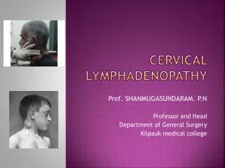
Tuberculosis lymphadenopathy PresentingSignsand Symptoms • Cervical nodes most commonly involved • Usual course of lymph node disease is as follows: Firm, discrete nodes fluctuant nodes matted together skin breakdown, abscesses, chronic sinuses healing and scarring
Diagnostics • Fine-needle aspiration of the involved lymph node • Extra-thoracic lymph node aspiration • Positive smears for acid-fast bacilli on fine-needle aspirates of the involved lymph nodes (high rate in HIV patients) • In smear-negative pulmonary TB, it is worthwhile aspirating extra-thoracic lymph nodes to confirm diagnosis of TB (80% positive)
TB abscess as part of immune reconstitution syndrome
Management and Treatment • Treatment should be started following the national TB Guidelines. • For further details, see Part A Module 2, Session 3.
Unique features, Caveats • One of the most common forms of extra-pulmonary TB in HIV patients • Fluctuant cervical nodes that develop over weeks to months without significant inflammation or tenderness suggest infection with M. tuberculosis, atypical mycobacteria, or scratch disease (Bartonella henselae). • In severe immunocompromised patients, tuberculosis lymphadenopathy may be acute and resemble acute pyogenic lymphadenitis • Miliary TB is an important consideration in patients with generalized lymphadenopathy