Download
1 / 65
760 likes | 1.86k Vues
PROTEIN METABOLISM. DR AMINA TARIQ BIOCHEMISTRY. In healthy, well fed individuals, the input to the amino acid pool is balanced by the output, that is, the amount of amino acids contained in the pool is constant. The amino acid pool is said to be in a steady state.

E N D
PROTEIN METABOLISM DR AMINA TARIQ BIOCHEMISTRY
In healthy, well fed individuals, the input to the amino acid pool is balanced by the output, that is, the amount of amino acids contained in the pool is constant. The amino acid pool is said to be in a steady state.
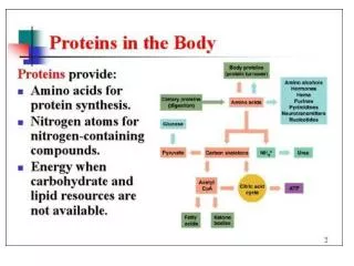
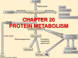
The "nitrogen or amino acid pool" is a grand mixture of amino acids available in the cell derived from dietary sources or the degradation of protein. Since proteins and amino acids are not stored in the body, there is a constant turnover of protein.
Some protein is constantly being synthesized while other protein is being degraded. For example, liver and plasma proteins have a half-life of 180 days or more, while enzymes and hormones may be recycled in a matter of minutes or hours.
Nitrogen Balance • nitrogen balance is achieved by a healthy person when the dietary intake is balanced by the excretion of urea wastes. If nitrogen excretion is greater than the nitrogen content of the diet, the person is said to be in negative nitrogen balance. This is usually interpreted as an indication of tissue destruction.
If the nitrogen excretion is less than the content of the diet, a positive nitrogen balance indicates the formation of protein.
Protein turnover • The recommended minimal protein intake required to achieve nitrogen balance in healthy adults is about 50g per day, although in developed countries many people may eat double this amount. This compares with an average daily protein turnover of about 250g per day.
Human proteins have very different lifetimes. Total body protein is about 11 kg, but about 25% of this is collagen, which is metabolically inert.
A typical muscle protein might survive for three weeks, but many liver enzymes turn over in a couple of days. Some regulatory enzymes have half-lives measured in hours or minutes. The majority of the amino acids released during protein degradation are promptly re-incorporated into fresh proteins.
Net protein synthesis accounts for less than one third of the dietary amino acid intake, even in rapidly growing children consuming a minimal diet..
Most of the ingested protein is ultimately oxidized to provide energy, and the surplus nitrogen is excreted, a little as ammonia but mostly as urea.
Soluble intracellular proteins are tagged for destruction by attaching ubiquitin, a low molecular weight protein marker. They are then degraded in proteasomes to short peptides
Transport of proteins • Amino acid uptake from the gut lumen into enterocytes is driven by the sodium gradient. • There is a relatively high sodium concentration in the gut and a low concentration in the enterocytes, as a result of the sodium pump in the basolateral membrane.
The concentration of free amino acids in the extracellular fluids is significantly lower than that within the cells of the body. • This concentration gradient is maintained because active transport systems, driven by the hydrolysis of ATP, are required for movement of amino acids from the extracellular space into cells.
At least seven different transport systems are known that have overlapping specificities for different amino acids
The small intestine and the proximal tubule of the kidney have common transport systems for amino acid uptake; • therefore, a defect in any one of these systems results in an inability to absorb particular amino acids into the gut and into the kidney tubules
For example, one system is responsible for the uptake of cystine and the dibasic amino acids, ornithine, arginine, and lysine (represented as “COAL”). • In the inherited disorder cystinuria, this carrier system is defective, and all four amino acids appear in the urine
Cystinuria occurs at a frequency of 1 in 7,000 individuals, making it one of the most common inherited diseases, and the most common genetic error of amino acid transport. • The disease expresses itself clinically by the precipitation of cystine to form kidney stones (calculi), which can block the urinary tract. • Oral hydration is an important part of treatment for this disorder.
Function • GGT is present in the cell membranes of many tissues, including the kidneys, bile duct, pancreas, gallbladder, spleen, heart, brain, and seminal vesicles • It is involved in the transfer of amino acids across the cellular membrane.
It is also involved in glutathione metabolism by transferring the glutamyl moiety to a variety of acceptor molecules including water, certain L-amino acids, and peptides
This general reaction is: (5-L-glutamyl)-peptide + an amino acid peptide + 5-L-glutamyl amino acid
Removal of Nitrogen • Removal of nitrogen from amino acids is essential for producing energy from amino acids. • It is an obligatory step in the catabolism of amino acids. • Once removed this N can be incorporated into other compounds or excreted.
STEPS • Mechanism of action • ALT/AST- substrate specificity • Pyridoxal phosphate • Equilibrium constant • Diagnostic value
Glutamate and glutamine play especially critical roles in nitrogen metabolism, acting as a kind of general collection point for amino groups • In the cytosol of hepatocytes, amino groups from most amino acids are transferred to -ketoglutarate to form glutamate, which enters mitochondria and gives up its amino group to form NH4
Excess ammonia generated in most other tissues is converted to the amide nitrogen of glutamine,which passes to the liver, then into liver mitochondria • Glutamine or glutamate or both are present in higher concentrations than other amino acids in most tissues
In skeletal muscle, excess amino groups are generally transferred to pyruvate to form alanine, another important molecule in the transport of amino groups to the liver.
Two reactions are important: • Transamination • Oxidative deamination
TRANSAMINATION REACTIONS • Most common amino acids can be converted into the corresponding keto acid by transamination. • This reaction swoops the amino group from one amino acid to a different keto acid, thereby generating a new pairing of amino acid and keto acid. • There is no overall loss or gain of nitrogen from the system.
One of the two pairs is almost invariably glutamate and its corresponding keto acid alpha ketoglutarate • These reactions are catalyzed by TRANSAMINASES(aminotransferases) • All transaminases require pyridoxal phosphate (derived from vitamin b6) as a coenzyme.
These enzymes are found in the cytosol and mitochondria of cells- • Liver, kidney ,intestine and muscle. • Alanineaminotransferase • Aspartateaminotransferase
Mechanism of action • The substrates bind to the active centre one at a time, and the function of the pyridoxal phosphate is to act as a temporary store of amino groups until the next substrate comes along. • In the process the pyridoxal phosphate is converted into pyridoxamine phosphate, and then back again.
AlanineTransaminase • This very active enzyme is known as alanineaminotransferaseand exists in mitochondrial and cytosolic variants. • The detailed iso-enzyme pattern is tissue-specific. • It escapes in large amounts from dead or dying tissues and GPT (glutamate pyruvateTransaminase) may be measured in blood samples for medical diagnostic purposes.
Alanine is the principal amino acid released from muscle tissue during starvation. It is an important substrate for hepatic gluconeogenesis • and Alaninetransamination is required for the proper maintenance of fasting blood glucose concentrations.
The enzyme catalyzes the transfer of the amino group of alanine to α-ketoglutarate, resulting in the formation of pyruvate and glutamate.
AspartateTransaminase • This enzyme known as aspartateaminotransferaseand is one of the most active enzymes in the cell. • Also called glutamate oxaloacetatetransaminase • It exists in mitochondrial and cytosolic variants, and the detailed iso-enzyme pattern is tissue-specific.
It escapes in large amounts from dead or dying tissues and enters the bloodstream, so AST is often measured in blood samples for medical diagnostic purposes.
AST is an exception to the rule that aminotransferases funnel amino groups to form glutamate. • During amino acid catabolism, AST transfers amino groups from glutamate to oxaloacetate, forming aspartate, which is used as a source of nitrogen in the urea cycle
Glutamate and aspartate are each required for separate but essential steps in the urea cycle, which is responsible for ammonia detoxification and nitrogen excretion. • The free movement of nitrogen between the glutamate and aspartate pools is an important balancing process that is vital for normal cellular metabolism.
Assays for Tissue Damage • Alanineaminotransferase (ALT; also called glutamate-pyruvatetransaminase, GPT) and aspartateaminotransferase (AST; also called glutamateoxaloacetatetransaminase, GOT) are important in the diagnosis of heart and liver damage caused by heart attack, drug toxicity, or infection.
After a heart attack, a variety of enzymes, including these aminotransferases,leak from the injured heart cells into the bloodstream. Measurements of the blood serum concentrations of the two aminotransferases by the SGPT and SGOT tests (S for serum)—and of another enzyme, creatinekinase, by the SCK test—can provide information about the severity of the damage.
Creatinekinase is the first heart enzyme to appear in the blood after a heart attack; it also disappears quickly from the blood. GOT is the next to appear, and GPT follows later. • Lactate dehydrogenase also leaks from injured or anaerobic heart muscle.