Download
1 / 68
840 likes | 1.9k Vues
Acute Pancreatitis. Umit Akyuz , MD Gastroenterology Department,Yeditepe University Istanbul. Acute Pancreatitis - Objectives. Discuss basic physiology Etiology Clinical Presentation Diagnosis Prognosis Management Complications. Pancreatic Physiology.

E N D
Acute Pancreatitis UmitAkyuz, MD GastroenterologyDepartment,YeditepeUniversityIstanbul
Acute Pancreatitis - Objectives • Discuss basic physiology • Etiology • Clinical Presentation • Diagnosis • Prognosis • Management • Complications
Pancreatic Physiology • (1) Production of bicarbonate-rich fluid to neutralize gastric fluid in the duodenum – duct cells primarily (CFTR gene = chloride / bicarbonate channel) • (2) Synthesis of digestive enzymes – acinar cells • (3) Insulin production = islet cells
Pancreatic Physiology • Most of the pancreas is made of acinar cells • The duct cells connect the acinar cells to the duodenum and as noted produces a bicarbonate rich fluid to “wash” the enzymes into the duodenum (3000cc/day) • The islet cells function independently but need remain in close proximity to the acinar cells to maintain normal gene expression
Pancreatic Physiology • Acinar enzymes are produced as zymogens • Zymogens are proenzymes that require cleavage of a peptide by trypsin to become active • Trypsinogen (zymogen of trypsin) is activated by the intestinal brush boarder enzyme enterokinase or by another trypsin molecule
Acute Pancreatitis – Epidemiology • >200,000 Hospital Admissions / Year • 20% have a severe course • 10-30% mortality for this group, which has not significantly changed during the past few decades despite improvement in critical care and other interventions
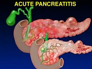
Acute Pancreatitis – Pathophysiology • An acute inflammatory process within the pancreas with variable involvement of localized tissues and remote organ systems • It can range from Mild Severe with MOSF, necrosis, abscess, or a myriad of other complications • Vigorous immune response • By the time pancreatitis is recognized clinically the inflammatory consequences dominate the clinical picture
Acute Pancreatitis • The vast majority of protection centers on trypsin activation (or lack thereof) • (1) preventing trypsinogen activation (2) inactivating trypsin (3) “sweeping” trypsin out of the pancreas
Acute Pancreatitis Etiology
Etiology • Alcohol (40%) • Mechanism not fully understood • Not all alcoholics get pancreatitis (only about 15%) • This suggests a subset of the population predisposed to pancreatitis, with alcohol acting more as a co-precipitant
Alcohol related pancreatitis - CASE • 26M presents for pseudocyst drainage – four weeks after severe pancreatitis • Has had “abdominal symptoms” since childhood • Does admit to heavy alcohol use as young adult • Strong family history of alcoholism and pancreatitis • Family history reveals a possible autosomal dominant abnormality • ERCP reveals chronic pancreatitis
Alcohol related pancreatitis - CASE • Answer: • A mutation (PRSS1) was found that alters the protein structure of trypsin producing a constant ON trypsin (auto-activation within the pancreas), resulting in frequent bouts of pancreatitis.
Etiology • Gallstones (35%) • Gallstone pancreatitis risk is highest among patients with small GS < 5mm and with microlithiasis • GS pancreatitis risk is also increased in white women > 60 yrs
Etiology – Drugs and Toxins (5%) • Azathioprine • Cimetidine • Estrogens • Enalapril • Erythromycin • Furosemide • Multiple HIV medications • Scorpion Bites • Sulfonamides • Thiazides • TMP/SMX
Etiology – Trauma • Blunt Trauma • Automobile • Bicycle handlebar injuries • Abuse • Iatrogenic – ERCP (1-7%) • Likely secondary to contrast but also very operator dependant • Risk is also increased with Sphincter of Oddi manometry
Etiology – Multi-System Disease • Diabetic Ketoacidosis (10-15%) • Hemochromatosis • HUS • Hypercalcemia • Hyperparathyroidism • Hypertriglyceridemia • IBD • Malnutrition • Severe PUD • Renal Failure • SIRS • SLE and other connective tissue dissorders • Status-Post solid organ and BM transplant • Vasculitis
Etiology – Multi-System Disease • Cystic Fibrosis • 2-15% of patients • Ductal obstruction from thickened secetions
Etiology – Multi-System Disease • Malnutrition and Re-feeding • Anorexia Nervosa • Pancreatic acinar cells atrophy but true cause of pancreatitis unknown
Etiology – Infection • Ascaris • Campylobacter • CMV • Coxsackie B • EBV • Enterovirus • HIV/AIDS • Influenza • MAC • Measles • Mumps Rubella • Mycoplasma • Rubeola • Viral Hepatitis • Varicella
Etiology – Anatomical Anomalies • Pancreas Divisum • Failure of dorsal and ventral fusion (5-15% of population) • Annular Pancreas • Any Ductal Anomalies • Sphincter of Oddi dysfunction • Always consider a primary malignancy as a possible cause of new onset pancreatitis in older patients without other obvious risk factors
Etiology – Genetic • CFTR heterozygote – “atypical” CF • Other CFTR mutations – found in many adult patients with more severe forms of pancreatitis (we can test for a few hundred of the more than 1200 different mutations) • SPINK1 mutations – most common cause of familial pancreatitis; causes a predisposition for pancreatitis (other trigger is usually present) • PRSS1 gene – Self-activating trypsin
Etiology - Autoimmune • Primarily a pancreatic disorder but . . . • Can be associated with other diseases of presumed autoimmune etiology including sclerosing cholangitis, primary biliary cirrhosis, retroperitoneal fibrosis, rheumatoid arthritis, sarcoidosis, and Sjögren's syndrome - some authors have proposed that AIP represents a systemic autoimmune disease
Etiology - Autoimmune • (1) Diagnostic histology - dense lymphoplasmacytic infiltrate of the pancreatic parenchyma with secondary fibrosis • (2) Characteristic imaging with elevated IgG4 • focal or diffuse enlargement of the pancreas that is often sausage-shaped • minimal pancreatic stranding • calcifications or peripancreatic fluid • uniform narrowing of the main pancreatic duct • rim-like enhancement of the pancreatic head • (3) Response to steroid therapy
Etiology – Idiopathic • Still accounts for ~20% of cases
Etiology – Idiopathic • Experts suggest that idiopathic pancreatitis should account for no more than 5-10% of the total cases, yet the broadly quoted percentage in the literature at this time in the US is currently 20-25%.
Acute Pancreatitis Clinical Presentation
Clinical Presentation • Clinical • Continuous mid-epigastric / peri-umbilical abdominal pain Radiating to back, lower abdomen or chest • Emesis • Fever • Aggravated by eating • Progressive • Restless and uncomfortable
Clinical Presentation • More severe cases • Jaundice • Ascites • Pleural effusions – generally left-sided • Cullen’s sign – bluish peri-umbilical discoloration • Grey Turner’s sign – bluish discoloration of the flanks
Diagnosis – Initial work-up • GS history • Drug intake • Family History • Alcohol intake • Viral exposures • Lipase • LFTs • GB US
Diagnosis – Follow up investigations • Fasting liver profile • Fasting plasma calcium – when well • Viral titers as indicated by clinical presentation • GB US – consider repeating • MRCP • CT pancreas (pancreas protocol)
Diagnosis – Further investigations • Appropriate for recurrent idiopathic acute pancreatitis • GB US • EUS / ERCP – biliary and pancreatic cytology • Autoimmune markers • Sphincter of Oddi manometry • Functional testing / History to screen for sequela of chronic pancreatitis
Diagnosis – Amylase • Elevates within HOURS and can remain elevated for 4-5 days • High specificity when using levels >3x normal • Many false positives (see next slide) • Most specific = pancreatic isoamylase (fractionated amylase)
Pancreatic Source Biliary obstruction Bowel obstruction Perforated ulcer Appendicitis Mesenteric ischemia Peritonitis Salivary Parotitis DKA Anorexia Fallopian tube Malignancies Unknown Source Renal failure Head trauma Burns Postoperative Diagnosis – Amylase Elevation
Diagnosis – Lipase • The preferred test for diagnosis • Begins to increase 4-8H after onset of symptoms and peaks at 24H • Remains elevated for days • Sensitivity 86-100% and Specificity 60-99% • >3X normal S&S ~100%
Diagnosis • Elevated ALT > 3x normal (in a non-alcoholic) has a positive predictive value of 95% for GS pancreatitis
Diagnosis – Imaging • CT • Excellent pancreas imaging • Recommended in all patients with persisting organ failure, sepsis or deterioration in clinical status (6-10 days after admission) • Search for necrosis – will be present at least 4 days after onset of symptoms; if ordered too early it will underestimate severity • Follow-up months after presentation as clinically warranted for CT severity index of >3
Diagnosis - Imaging • ERCP / EUS • Diagnostic and Therapeutic • Can see and treat: • Ductal dilatation • Strictures • Filling defects / GS • Masses / Biopsy
Diagnosis – Imaging • ERCP indications (should be done in the first 72hr) • GS etiology with severe pancreatitis – needs sphincterotomy • Cholangitis • Jaundice • Dilated CBD • If no GS found sphincterotomy is indicated anyway • Poor surgical candidate for laparoscopic cholecystectomy • Clinical course not improving sufficiently to allow timely laparoscopic cholecystectomy and intraoperative cholangiogram • Pregnant patient • Uncertainty regarding biliary etiology of pancreatitis
Acute Pancreatitis Prognosis
Prognosis – Ranson’s (Severe > 3) • Ranson’s Score • 5 on Admission • Age > 55 y • Glucose >200 • WBC > 16000 • LDH > 350 • ALT > 250 • 6 after 48 hours from presentation • Hct > 10% decrease • Calcium < 8 • Base Deficit > 4 • BUN > 5 • Fluid Sequestration > 6L • PaO2 < 60
Prognosis – Atlanta Criteria • Severe Acute Pancreatitis • Early Prognostic Signs • Ranson signs ≥3 • APACHE-II score ≥8 • Organ Failure and/or Local Complications (persist >48 hr after admission) • Necrosis • Abscess • Pseudocyst • Organ Failure as Defined by Atlanta Symposium • Shock–systolic pressure <90 mmH • PaO2 ≤60 mmHg • Creatinine >2.0 mg/L after rehydration • Gastrointestinal bleeding >500 cc/24 h
Prognosis – CT Severity Index • CT Grade • Normal 0 points • Focal or diffuse enlargement 1 point • Intrinsic change or fat stranding 2 points • Single ill-defined fluid collection 3 points • Multiple collections of fluid or gas 4 points • Necrosis Score • None 0 points • 1/3 of pancreas 2 points • 1/2 of pancreas 4 points • > 1/2 of pancrease 6 points • Severe = Score > 6 (CT Grade + Necrosis)
Prognosis – CRP • Santorini consensus and the World Association guidelines recommend a cut off of 10 - 15 mg/dl • This is checked 48 hours after admission
Management • All patients with biliary pancreatitis should undergo definitive treatment of gallstones during the same hospital admission, unless a clear plan has been made for definitive treatment within the next two weeks • Delay exposes the patient to the risk of potentially fatal recurrent acute pancreatitis • Surgery should be delayed in severe pancreatitis and ERCP is preferred
Management • Mainly supportive • Hydration, pain relief, and pancreatic rest • NPO – to decrease pancreatic secretion • Remember stress ulcer prophylaxis always • Look for complications!!! • No magic bullet – antiproteases, octreotide (antisecretory) or lexiafant (anti-inflammatory) have all been disappointing in large trials
Management - Antibiotics • Infected necrosis has significant mortality (40%) • No consensus at this time for prophylactic antibiotics but the literature leans toward using them in severe pancreatitis with necrosis • Imipenum, cefuroxime, ceftazidime + amikacin + metro, Ofloxacin + metro, cipro + metro your choice • If used, it should be given no longer than 7 to 14 days • Gut decontamination is not recommended
Management - Feedings • Enteral nutrition is preferred • There is a push for nasojejunal feeds however nasogastric feeds have been shown to be effective in 80% of cases¶ • NGTs should be used with caution in patients with AMS however • More risk with TPN / IL but if cannot feed enterally >5 days may be needed ¶ Eatock FC. Nasogastric feeding in severe acute pancreatitis. Radiology 1994: 193, 297-306.
Management – Necrosis • All severe pancreatitis should be managed in the ICU or Stepdown Unit • FNA recommended when >30% necrosis or clinical suspicion of sepsis for a culture 7-14 days after onset, if gas present assume infected necrosis and treat • Infection generally requires debridement (surgical or IR)
Management – Pain • Morphine not ideal but can still be used – it can theoretically worsen symptoms by increasing spasm of the Sphincter of Oddi • Demerol is a good opiate agonist • Hydromorphone is also an excellent option • PCA is generally preferred in the beginning • Always use the gut if you can to transition off IV pain meds • Don’t forget aggressive bowel care