Download
1 / 60
610 likes | 920 Vues
Metastatic Mucinous Adenocarcinoma (MAC). Sherif R.Z. Abdel-Misih, MD Assistant Professor of Surgery Division of Surgical Oncology The Ohio State University Medical Center/James Cancer Hospital. Metastatic MAC. Objectives Discuss Colorectal MAC/Implications

E N D
Metastatic Mucinous Adenocarcinoma (MAC) Sherif R.Z. Abdel-Misih, MD Assistant Professor of Surgery Division of Surgical Oncology The Ohio State University Medical Center/James Cancer Hospital
Metastatic MAC Objectives Discuss Colorectal MAC/Implications Discuss Appendiceal Mucinous Neoplasms Discuss Role of Cytoreductive surgery/HIPEC
Colorectal Adenocarcinoma Colorectal Cancer remains 3rd most common cancer in U.S. with associated mortality Estimated 142,570 new colorectal cancer cases in 2010 Estimated 51,370 deaths in 2010 Jemal A et al. CA Cancer J Clin 2010: 60(5):277-300
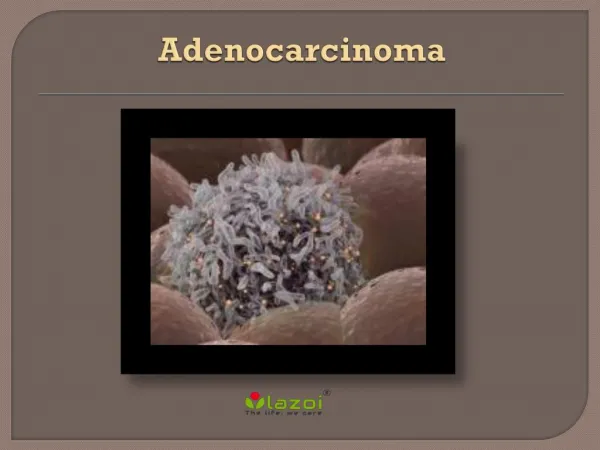
Adenocarinoma 3 distinguishing types of Colorectal AdenoCa Mucinous AdenoCa Non-Mucinous Signet Ring (SR)
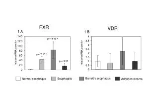
MAC Clinical implications of Mucinous pathology unclear based on literature to date Most studies are retrospective series without concordant findings
SEER DB Colon Ca SEER DB (1992-2000) 164,628 patients evaluated from 1991-2000 Only 10.3% with Mucinous Histology Similar clinical characteristics Kang H. Dis Colon Rectum 2005: 48(6):1161-8.
Incidence rate per 100,000 Clinicopathologic Characteristis
Survival Comparison Worse 5yr. Overall Survival for MAC vs. AC But not stage for stage Signet ring cell clearly conferred worst survival HR of 1, 1.01, and 1.372 for AC, MAC, Signet ring, respectively
SEER DB conclusions MAC has differing clinical characteristics Right sided primary more commonly Mucin production Slightly more advanced staged disease Increase Node + Disease Peritoneal Disease MAC has worse 5yr OS compared to adenocarcinoma, but similar survival patterns stage for stage based on SEER ?Worse prognosis: SR > MAC ≥ AC
MAC (Song W) Retrospective Chinese study with 2006 pts with CRC b/w 1994-2007 144 (7.2%) MAC 1837 (91.2%) AdenoCa 25 (1.2%) SR Younger patients (Mean age); p<0.001 54.2 MAC 58.7 AdenoCa 40.4 SR Song W. Chinese Medical Journal 2009: 122(13): 1486-1491
Clinicopathologic Differences Tumor Size; p<0.001 5.52cm MAC 5.52cm SR 4.62cm AdenoCa MAC/SR Increased LN involvement, peritoneal dz, serosal infilration/organ invasion (p<0.0001)
Predictors of Survival Factors affecting survival Stage, Histology, age, tumor site, depth of invasion Mucinous/SR histology were negative predictors of survival (p<0.01)
Overall Survival Statistically significant difference in median OS for all pts (p<0.01) MAC 67.7 mos Adenoca 151 mos SR 32.6 mos 5yr OS (p<0.01) MAC 51% Adenoca 69% SR 27% Median OS in all pts
Survival after Curative Rsxn Median OS after curative resection MAC 135 mos Adenoca 151 mos SR 41.1 mos P<0.003 5yr OS MAC 62% Adenoca 70% SR 28% (p<0.01) Median OS in pts after curative resection
Survival (Stage-specific II/III) No difference in Stage I or IV Comparing OS Stage II/III MAC 135 mos AdenoCa 149 mos SR 33 mos P<0.001
Colorectal MAC Conclusions Not well studied to date Prognosis for mucinous histology may be equivalent or slightly worse overall than non-mucinous counterpart Signet-Cell clearly worse prognosis
Metastatic Colorectal MAC Tx Improved systemic therapy (Cytotoxic, Biologics) has increased treatment options Prolonged patient survivals May induce tumor shrinkage → ↑ Surgical resectability Surgical resection best option in selected patients with alternative regional therapy (i.e. ablation, SIRT, TACE, HAIP, IRE)
Colorectal MAC Metastasectomy Liver metastases common ( ≈ 50%) and often only site Chemotx has allowed for resection in up to 20% of pts with colorectal liver metastases (CLM) previously deemed ‘unresectable’ with 5yr survivals exceeding 50% (Choti, Ann Surg 2002) Previous surgical contraindications no longer substantiated Bilobar disease >4 lesions Extrahepatic disease (EHD) Lesions >5cm Metachronous disease Metastasectomy + Chemotherapy is best option for prolonged survival/cure
Metastasectomy (EHD) de Haas/Adam et al.(Dig Surg 2008. 25(6)) 5-yr survivals of over 30% in pts with concomittant lung metastases after resection of liver/lung dz Aggressive therapy may be considered for those that are cytoreducible and responsive to systemic therapy
Colorectal MAC Multidisciplinary approach important Surgery still essential in management of primary and metastatic colorectal MAC Advocate aggressive surgical/medical management of colorectal adenocarcinoma (Mucinous/Non-Mucinous)
Appendiceal Mucinous Neoplasms Account for <3% of all appendectomy specimens and <0.5% of all GI tract malignancies Unlike colorectal MAC, <5% exhibit lymph node disease and often loco-regional disease Intestinal type histology has much higher association with LN disease Association between Appendiceal mucinous neoplasms and pseudomyxoma peritonei (PMP)
Appendiceal Mucinous Neoplasms Different and more favorable natural hx than colorectal MAC Challenging clinicopathologic disease Pathologic classification schema continue to change without general consensus Without pathologic consensus, reported literature variable Disease biology/course not well understood and hence treatment consensus lacking
Appendiceal Mucinous Neoplasms Pathology classifications Misdraji (Misdraji J. Am J Surg Path: 2003, 27(8)) Low grade Appendiceal Mucinous Neoplasm (LAMN) Mucinous Adenocarcinoma Ronnett (Ronnett BM. Am J Surg Pathol 1995; 19(12)) DPAM (Disseminated peritoneal adenomucinosis) PMCA (Peritoneal mucinous carcinomatosis) Pai&Longacre (Pai RK. Adv Anat Pathol 2005. 12(6)) Adenoma Mucinous tumor, uncertain malignant potential Mucinous tumor, low malignant potential Adenocarcinoma PMP considered by many a low grade malignancy due to its malignant, but indolent behavior
Pathology implications Low grade histology is applied to those lesions composed of mucin with proliferative mucinous epithelium without cytologic atypia High grade histology applies to lesions with abundant epithelium with noted cytologic atypia
Appendiceal Neoplasms: SEER DB 2514 pts from 1973-2001 in SEER DB analyzed Histologic classification into 5 groups Mucinous (951) Adenocarcinoma (646) Carcinoid (435) Signet Ring (113) Goblet (369) Analyzed clinicopathologic characteristics and survivals by histology McGory M. Dis Colon Rectum. 2005;48(12):2264-71.
Clinicopathologic Characteristics Comparable groups except for tumor size and disease stage at presentation Increased size primary tumors at presentation compared to other histology Present with advanced stage disease (Adeno < Mucinous < SR)
Appendical MAC Survival Improved 5yr OS stage for stage compared to non-mucinous Appendiceal AdenoCa Regional Dz: 54% vs 37% (p<0.001) Distant Dz: 32% vs. 11% (p<0.05)
Pai/Longacre Pai/Longacre examined 116 pts with varied pathology and proposed a prognostic pathhologically based schema Pathologic features associated with 4 groups Pai RK. Am J Surg Path 2009. 33(10):1425-1439.
Pai - Clinical Features Clinical characteristics by groups/pathology Often present with or misdiagnosed as appendicitis Advanced disease may demonstrate mass (?Abscess) on imaging
OS & DFS by Pathology OS and DFS reflects indolent nature of disease based on pathologic staging
Predictors of Survival/Prognosis Advanced stage (group) and regional disease associated with more aggessive biology/worse prognosis Univariate predictors for OS extra-appendiceal mucin (p=0.01) HG cytology (p<0.0001) complex architecture (p<0.001) invasion (p<0.001) Multivariate predictors for decreased OS/DFS extra-appendiceal mucin (p=0.006) HG cytology (P=0.001)
Appendiceal Mucinous Neoplasm management? No consensus Extensive pathologic review of available pathology important to treatment planning Previous/Current oncology dogma suggests right hemicolectomy ? Hyperthermic intraperitoneal chemotherapy (HIPEC)
Right Hemicolectomy?(Gonzalez-Moreno) Gonzalez-Moreno et al. examined 501pts from 1983-2000 Aimed to elucidate need/benefit of right hemicolectomy in management of appendiceal neoplasms Gonzalez-Moreno S. Br Journal of Surgery 2004; 91:304-311
Survival (Gonzalez-Moreno) Median OS all pts was 13 yrs Median OS based on histology Mucinous 13 yrs – Better Prognosis Intestinal 5 yrs Not statistically significant
Overall Survival By univariate analysis, Median overall survival based on surgical procedure was significant, p<0.001 18 years appendectomy 10 years R hemicolectomy 4 years for none (due to inability to attain complete cytoreduction) Procedure was not independent predictor on MV analysis (p=0.258)
Survival (Surgery/HIPEC) Improved Median OS with appendectomy alone/Right colectomy w/ HIPEC compared to Right colectomy 16yrs Appy or R colectomy w/ HIPEC 9 yrs R colectomy w/o HIPEC P=0.007
Survival (Node Disease) 120/501(24%) pts w/ nodal assessment Only 25pts (5%) had (+)Ln Disease 25/120 (21%) LN+ pts with LN assessment Lymph node Dz by histology (p<0.001) Intestinal histology 66.7% Mucinous histology 4.2%
R Hemicolectomy?? Conclusions Not indicated in all patients Due to low risk for nodal disease and no positive impact on survival Selective use in pts Aggressive Pathology/Invasive Disease + LN disease + Appendiceal resection margin When necessary to clear primary tumor or achieve complete cytoreduction Nonmucinous appendiceal histology due to higher propensity for Nodal disease
Peritoneal Carcinomatosis (PC) PC is typically late manifestation of advanced tumor stage, progression, or recurrence of various cancers Historically poor prognosis and outcomes Posed significant treatment challenges for surgeons and medical oncologists Lack of good treatment options
PC Historical treatment Systemic chemotherapy and radiation therapy offered little benefit if any Surgical treatment was palliative in nature Enteric Bypass procedures ± Enterostomy Ovarian cancer was first tumor type treated with CRS in early 1980’s Mid-Late 1980’s, investigation into CRS/HIPEC by Paul Sugarbaker (WCI) with some encouraging results - spurred further study
CRS/HIPEC 2 Major components Cytoreductive Surgery Addresses gross peritoneal tumor burden Goal of surgery is to eliminate all gross disease (<2.5mm) Hyperthermic Intraperitoneal Chemotherapy Addresses microscopic peritoneal disease after CRS using intraperitoneal perfusion
HIPEC Hyperthermia More toxic to cancer cells than normal cells; more pronounced with increased vascularity Potentiates cytotoxic effects of chemotherapy Direct effect on tumor tissue to soften the tissue and decreases interstitial pressure, hence improving drug penetration
Cytoreductive Surgery Major Surgery Eliminate all gross tumor burden Peritoneal Dz may requires peritonectomy Multivisceral(Organ) Resection sometimes necessary
Cytoreduction Peritoneal Surface Malignancy Group determined 8 factors predictive of CCR ECOG≤2 No evidence of extra-abdominal disease ≤3 small, resectable hepatic metastases No biliary obstruction No ureteral obstruction No Bowel obstruction at > 1 site SB involvement: Disease within mesentery with several sites of partial obstruction Small volume disease in lesser omentum Esquivel J. Ann Surg Oncol 2007. 14: 128-133
HIPEC (Verwaal et al.) Verwaal et al. demonstrated survival advantage for patients with PC (colorectal) Median survival of 22.3 months in patients receiving CRS/HIPEC/Standard Chemotherapy Median Survival of 12.6 months in patients receiving chemotherapy(5-FU/Leucovorin) with or without palliative surgery (p=0.032) Verwaal V at al. JCO 2003. 21(20): 3737-3743
Cytoreductive Surgery (Glehen) Major determinant of survival Glehen et al. in multi-institutional study of 506pts with CRS/HIPEC Overall median survival was 19.2 months Complete CRS/HIPEC median survival 32.4 months Incomplete cytoreduction survival 8.4 months P<0.0001 Glehen et al. JCO 2004. 22(16): 3284-3292
CRS/HIPEC complications Major morbidity associated with CRS and HIPEC Morbidity 22.9% Mortality 4% Glehen et al. JCO 2004. 22(16): 3284-3292