Download
1 / 15
150 likes | 343 Vues
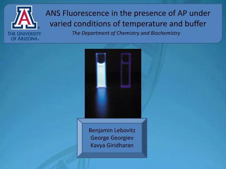
ANS Fluorescence in the presence of AP under varied conditions of temperature and buffer. The Department of Chemistry and Biochemistry. Benjamin Lebovitz George Georgiev Kavya Giridharan. Fluorescence Spectroscopy. Flourophores are molecules which can respond distinctly to light

E N D
ANS Fluorescence in the presence of AP under varied conditions of temperature and buffer The Department of Chemistry and Biochemistry Benjamin Lebovitz George Georgiev Kavya Giridharan
FluorescenceSpectroscopy • Flourophores are molecules which can respond distinctly to light • Excitation • Emission • ANS Excitation: 380nm
8-Anilinonaphthalene-1-sulfonate(ANS) • Fluorescent probe which binds to hydrophobic clusters in Alkaline Phosphatase enzyme • Hydrophobic interactions • Coulomb interaction Sulfonate anion • Shift in ANS emission peak upon binding to protein • Flourophore must be removed from H2O in order to fluoresce Non-polaranilinophthalein ANS
E. Coli 12 Alkaline Phosphatase • A homodimeric enzyme containing α-helical motifs which enclose parallel beta strands mostly consisting of hydrophobic residues • Structure is stabilized by two disulfide bonds present within each monomer • The disulfide bonds near protein surface and are susceptible to disruption when in the presence of reducing agents (TCEP)
TCEP • Powerful reducing agent • Irreversible cleavage of disulfide bonds destabilizes structure • Protein cannot refold • Disulfide bonds (red) • Active site Mg+2(magenta) • Active site Zn+2(blue) • Active site Ser residue (yellow)
Methods and Materials • Photon Technology International Fluorimeter • AP solution in 10 μM Tris 7.4, 5 μM MgSO4 • AP solution in 10 μM Tris 7.4, 5 μM MgSO4, 5 μM ZnCl2 • Buffer with 10 μM Tris 10 mM MgSO4 pH 7.4 • Buffer after dialysis with 10 μM Tris 7.4, 5 μM MgSO4, 5 μM ZnCl2 • 252 μM ANS solution • 10 mM TCEP *concentrations of each species in cuvette was kept constant *total cuvette volume was kept constant (1500 μL) *75 mm slit width used
Methods and Materials • ANS baseline spectrums were run in both buffers, +/- TCEP • Fluorescence of ANS with AP in both buffers was measured • Fluorescence of ANS with AP and TCEP in both buffers was measured • Each solution’s fluorescence was measured in a spectrum of 420 to 620 nm. • Temperature was increased from 20 °C to 40 °C, 60 °C, 90 °C and cooled back to 20 °C with fluorescence of the solution measured at each point.
Variation of ANSFluorescence with change in temperature, buffer, and presence of TCEP 2 μM AP + 20.2 μM ANS in 10mM Tris (pH=7.4), 5mM MgSO4 buffer ` * Raman band has been subtracted form spectrums
Variation of ANSFluorescence with change in temperature, buffer, and presence of TCEP 2 μM AP + 20.2 μM ANS in 10mM Tris (pH=7.4), 5mM MgCl2, 5mM ZnCl2 buffer ` * Raman band has been subtracted form spectrums
Variation of ANSFluorescence with change in temperature, buffer, and presence of TCEP 2 μM AP + 700μM TCEP + 20.2 μM ANS in 10mM Tris (pH=7.4), 5mM MgSO4 * Raman band has been subtracted form spectrums
Variation of ANSFluorescence with change in temperature, buffer, and presence of TCEP 700μM TCEP + 20.2 μM ANS in 10mM Tris (pH=7.4), 5mM MgCl2, 5mM ZnCl2 buffer * Raman band has been subtracted form spectrums
Fluorescence Quenching • Loss of fluorescence due to solvent interaction with the ANS fluorophore. • Less quenching is observed when protein is folded because of decreased access of solvent to buried hydrophobic regions. • As a protein becomes denatured, its hydrophobic regions are exposed to water. Interaction between ANS and the solvent induces a lower observed fluorescence.
Conclusions • Less ANS fluorescence when protein is fully denatured due to quenching • SO4-2 anions in solution stabilizes AP structure better than Cl- • Reducing agent TCEP denatures protein and impedes AP refolding upon cooling • Disulfide bonds cannot reform
References • Hung, H. C., and G. G. Chang. 2001. Unfolding of Placental Alkaline Phosphatase. Biophysical Journal 81(6) 3456–3471. • R. W. Poe et al. 1993. Effects of Various Salts on the Steady-State Enzymatic Activity of E. coli Alkaline Phosphatase.Journal of Inorganic Biochemistry, Vol. 15, 173-18. • Matulis et al. 1999. 1-Anilino-8-Naphthalene Sulfonate as a Protein Conformational Tightening Agent. Biopolymers, Vol. 49, 451–458. • M. Bortolato et al. 1999. Alkaline Phosphatase Ion-Dependent Structure. Proteins, Vol 37, 310–318.