Download
1 / 34
340 likes | 479 Vues
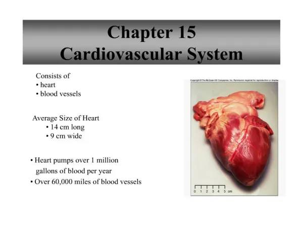
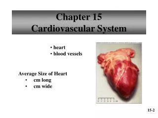
Cardiovascular System Chapter 13. Objectives: Identify structures and functions of the cardiovascular system. Trace the flow of blood through the body. Coverings of the Heart. Pericardium : (?) Visceral (?) pericardium (“epicardium” (?) ) Innermost layer Covers the heart

E N D
Cardiovascular SystemChapter 13 Objectives: Identify structures and functions of the cardiovascular system. Trace the flow of blood through the body.
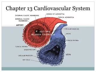
Coverings of the Heart • Pericardium: (?) • Visceral (?)pericardium (“epicardium”(?)) • Innermost layer • Covers the heart • Parietal (?)pericardium • Outer layer of visceral pericardium • Forms inner lining of fibrous pericardium • Fibrous (?)pericardium • Dense connective tissue attached to central portion of diaphragm, posterior of sternum, vertebral column, and large blood vessels attached to heart
Pericardial Cavity • Space between parietal and visceral pericardium layers • Filled with serous fluid to reduce friction between the membranes as the heart moves
Wall of the Heart • Epicardium • Outer layer • Corresponds to visceral pericardium • Protects heart by reducing friction • Myocardium • Thick, middle layer • Consists mostly of cardiac muscle tissue that pumps blood out of the heart chambers • Endocardium • Inner layer • Epithelium and connective tissue
Heart Chambers & Valves • ???? • Four hollow chambers: • Upper chambers = atria (atrium) • Lower chambers = ventricles • Atria • Thin walls • Receive blood returning to the heart • Ventricles • Receive blood from the atria • Contract to force blood out of the heart into the arteries
Heart Chambers & Valves, continued….. • Left atrium and ventricle are separated from the right atrium and ventricle by a solid, wall-like septum. • Atrioventricular valves – separate ??? • Tricuspid valve • on the right • “3 cusps” • Bicuspid valve (“mitral valve”) • on the left • “2 cusps” • Allows blood to flow ONLY from atrium to ventricular – no backflow
Heart Chambers & Valves, continued….. • Right atrium receives blood from: • Superior vena cava (???) • Inferior vena cava (???) • Coronary sinus – small vein that drains blood from the myocardium • Right ventricle • Thinner muscular wall than the left ventricle • Only has to pump blood to the lungs • Blood leaves the ventricle through the pulmonary valve, and enters the pulmonary trunk, which divides into the left and right pulmonary arteries.
Heart Chambers & Valves, continued….. • Left atrium receives blood from the lungs through 4 pulmonary veins. • Blood leaves the left ventricle through the aortic valve, into the aorta. • Ventricular wall is thicker than that of the right ventricle – has to pump blood to the rest of the body
Add to Your Labeled Diagrams • Color the areas that contain oxygenated blood red. • Color the areas that contain unoxygenated blood blue. (***WHAT COLOR IS UNOXYGENATED BLOOD, REALLY?) • Add arrows to indicate the direction of blood through the whole system. • Write in the areas each blood vessel is going to or coming from (i.e., lungs, body, heart muscle).
Blood Supply to the Heart • Coronary arteries • Supply oxygenated blood to the heart muscle • The first 2 branches off the aorta, right beside the aortic valve • These are tiny! • Cardiac veins • Return blood from the myocardium to the coronary sinus. • What is the coronary sinus?
Blood Vessels • Arteries → arterioles → capillaries → venules → veins → ??? • Gas exchange between the blood and body tissues occurs in which blood vessels? • Arteries are under relative high pressure. • Veins have valves that prevent a backflow of blood.
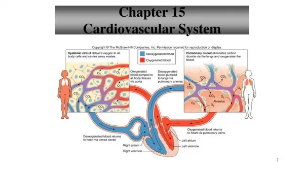
Paths of Circulation • Two major pathways: • Pulmonary circuit - lungs • Systemic circuit – body and heart • BOTH pathways have the 5 types of blood vessels. • The pulmonary circuit starts when blood is pumped out of the __________, and ends when blood returns to the _______. • The systemic circuit starts when blood is pumped out of the __________, and ends when blood returns to the _______.
Arterial System • Aorta • Largest diameter artery • Exits the left ventricle (1. ascending aorta) • Curves over the top and to the left of the heart (2. aortic arch) • 3. Descending aortagradually moves medially(?) until it lies directly in front of the vertebral column • Descending aorta ABOVE the diaphragm: 4. thoracic aorta • BELOW the diaphragm: 5. abdominal aorta
For this next part, you will need: • Handout to label as we go. • Add this chart to your notes *****:
Major Arteries of the AscendingAorta • Right and left coronary arteries
Major Arteries of the Aortic Arch • Brachiocephalic artery • Left common carotid artery • Left subclavian artery
Major Arteries of the Descending Aorta • Bronchial artery
Major Arteries of the Thoracic Aorta • Pericardial artery • Esophageal artery • Mediastinal artery • Posterior intercostal artery
Major Arteries of the Abdominal Aorta • Celiac artery ( gives rise to gastric, splenic, and hepatic arteries) • Phrenic artery • Superior mesenteric artery • Suprarenal arteries • Renal arteries • Gonadal arteries • Inferior mesenteric artery • Lumbar arteries • Middle sacral artery • Common iliac arteries
Arteries to the Neck, Head, and Brain • These branch off the subclavian and common carotid arteries. • Vertebral arteries • branch off the subclavian arteries • pass through the transverse foramina of the cervical vertebrae • Enter the skull through the foramen magnum • Supply blood to the vertebrae and their ligaments and muscles
Arteries to the Neck, Head, and Brain, continued….. • Thyrocervical arteries • short vessels that branch off the subclavian arteries • give off branches to the thyroid gland, parathyroid glands, larynx, trachea, esophagus, and pharynx
Arteries to the Neck, Head, and Brain, continued….. • Left and right common carotid arteries diverge into internal and external carotid arteries • External carotid artery goes up the side of the head giving off branches to structures in the neck, face, jaw, and base of the skull • Internal carotid artery follows a deeper course along the pharynx to the base of the skull • Provides the major blood supply to the brain
Arteries to the Shoulder and Upper Limb • After the subclavian artery pass between the clavicle and the 1st rib, it becomes the axillary artery, which then becomes the brachial artery. • A deep brachial artery branches off the brachial artery and curves around the humerus to supply the triceps brachii. • At the elbow, the brachial artery branches into radial and ulnar arteries. They rejoin at the wrist. • Which artery is used for taking a pulse?
Arteries to the Pelvis and Lower Limb • Common iliac arteries divide into internal and external arteries. • Internal supplies pelvic muscles and organs • External supplies lower limbs – becomes the femoral artery • Femoral artery supplies muscles and tissues of the thigh area. • As it approaches the knee, it becomes the popliteal artery, supplying the knee joint and muscles in the thigh and calf.
Arteries to the Pelvis and Lower Limb, continued….. • Popliteal artery branches into: • Anterior tibial artery • Posterior tibial artery • Anterior tibial artery: • Passes between tibia and fibula • Supplies skin and muscles in anterior and lateral parts of leg • Becomes the dorsalis pedis artery(???) • Posterior tibial artery: • Descends beneath the calf muscle • Supplies skin, muscles, and other tissues of the leg
Venous System • MOST veins follow closely along their same-named arteries. (example: brachial artery and brachial vein) • Notable differences: • Aorta vs. vena cava • Internal and external carotid arteries vs. internal and external jugular veins