Download
1 / 31
510 likes | 1.61k Vues
Ankylosing Spondylitis. M Handel 1 st Feb 2012. A Disease of Antiquity: Ankylosing Spondylitis . Amenhotep II (1439-1413 BC) 1 Rameses the Great (1298-1232 BC) 1. 1 Rheumatol Int. 2003; 23:1-5. . Ankylosing Spondylitis (AS).

E N D
Ankylosing Spondylitis M Handel 1st Feb 2012
A Disease of Antiquity:Ankylosing Spondylitis • Amenhotep II (1439-1413 BC)1 • Rameses the Great (1298-1232 BC)1 1Rheumatol Int. 2003; 23:1-5.
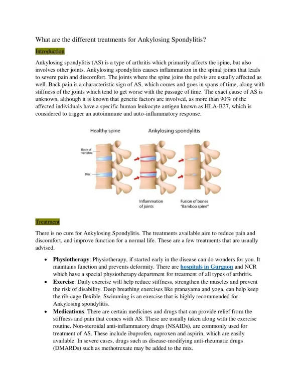
Ankylosing Spondylitis (AS) • AS is a chronic, progressive immune-mediated inflammatory disorder that results in ankylosis of the vertebral column and sacroiliac joints1 • The spine and sacroiliac joints are the common affected sites1 • Chronic spinal inflammation (spondylitis) can lead to fusion of vertebrae (ankylosis)1 1 Taurog JD. et al. Harrison‘s Principles of Internal Medicine, 13 th Ed. 1994: 1664-67.
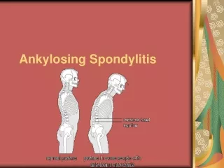
Ankylosing Spondylitis“Bamboo Spine” Repeated process of healing and bone formation leads to formation of syndesmophytes ‘bone bridges’ ACR Slide Collection on the Rheumatic Diseases; 3rd edition. 1994.
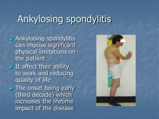
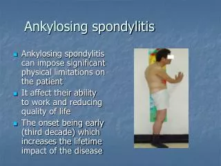
AS: A Debilitating Rheumatic Disease Over time, joints in the spine can fuse together and cause a fixed, bent-forward posture AS patients have an important impact on health care and non health-care resource utilization, resulting in a mean total cost (direct and productivity) of about $6700 to $9500/year/patient1 More than 30% of patients carry a heavy burden of disease and have a decreased QoL2 1Linden VD et al. Chapter 10. In: Firestein, Budd, Harris, McInnes, Ruddy and Sergent, eds. Kelley’s Textbook of Rheumatology: Spondyloarthropathies. 8th ed. Saunders Elsevier;2009:p.1171 2 Braun J & Sieper. J Rheumatology 2008;47:1738-40
AS (“Mis-”) Perceptions • 0.1-0.9%1,2 • “Rare” • “Not” a serious disease, functional limitation is mild • “Rarely shortens life” • Burden of disease significant in pain, sick leave, early retirement3,4,5 • Mortality figures parallel RA6,7,8 1 Sieper J et al. Ann Rheum Dis. 2002; 61 (suppl 3);iii8-18.2 Lawrence RC., Arthritis Rheum 1998; 41:778-99. 3 Zink A., et al., J Rheumatol 2000; 27:613-22. 4 Boonen A. Clin Exp Rheumatol. 2002;20(suppl 28):S23-S26. 5 Gran JT, et al. Br J Rheumatol. 1997;36:766-771. 6 Wolfe F., et al. Arthritis Rheum. 1994 Apr;37(4):481-94. 7 Myllykangas-Luosujarvi R, et al. Br J Rheumatol. 1998;37:688-690. 8 Khan MA, et al. J Rheumatol. 1981;8:86-90. 9 Braun J., Pincus T., Clin Exp Rheumatol. 2002; 20(6 Suppl 28):S16-22.
Epidemiology of AS • The incidence of AS may be underestimated due to unreported cases1 • HLA-B27 gene is associated with AS6 • Age of onset typically between 15 and 35 years1,2,3 • 2-3 times more frequent in men than in women6 1The Spondylitis Association of America. Available at: www.spondylitis.org. Accessed December 2,2004. 61(suppl 3);iii8–18.6Khan MA. Ann Intern Med. 2002;136:896–907.
SpA and HLA-B27 Khan MA. Ann Intern Med 2002;136(12):896-907
RA Age at Onset Distribution of AS and Rheumatoid Arthritis (RA) AS Percentage of Patients (%) Economically active individuals with a major impact on their ability to work1 1Barkham N et al. Rheumatology 2005;44:1277-1281 2ZinkA et al. Ann Rheum Dis 2001;60:199-206
Inflammation Disease activity Structural damage Syndesmophytes formation AS: Characteristic Pathologic Features • Chronic inflammation in: • Axial structures (sacroiliac joint, spine, anterior chest wall, shoulder and hip) • Possibly large peripheral joints, mainly at the lower limbs (oligoarthritis) • Entheses (enthesitis) • Bone formation particularly in the axial joints Sieper J. Arthritis Res Ther 2009;11:208 Elewaut D & Matucci MC. Rheumatology 2009;48:1029-1035
Inflammation Disease activity AS: Signs and Symptoms Axial manifestations: • Chronic low back pain • With or without buttock pain • Inflammatory characteristics: • Occurs at night (second part) • Sleep disturbance • Morning stiffness • Limited lumbar motion • Onset before age of 40 years MRI sacro-iliac joint Inflammatory back pain (IBP) = Characteristic symptom Sengupta R & Stone MA. Nat ClinPractRheumatol2007;3:496-503 Hultgren S et al. Scand J Rheumatol 2000;29:365-369 Linden VD et al. Chapter 10. In: Firestein, Budd, Harris, McInnes, Ruddy and Sergent, eds. Kelley’s Textbook of Rheumatology: Spondyloarthropathies. 8th ed. Saunders Elsevier;2009:p.1175
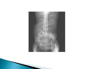
Structural damage Syndesmophytes formation AS: Structural Damage Most striking feature of AS = New bone formation in the spine with: • Spinal syndesmophytes • Ankylosis Both can be seen on conventional radiography X-ray showing syndesmophytes Even in patients with longer-standing disease, syndesmophytes are present in ~50% patients and a smaller percentage will develop ankylosis Bamboo spine and bilateral sacroiliitis SieperJ. Arthritis Res Ther 2009;11:208
AS: Signs and Symptoms Peripheral manifestations • Enthesitis • Peripheral arthritis • Dactylitis Up to 58% patients ever had arthritis1 Much smaller number of patients2 50% patients with enthesitis1 1Cruyssen BV et al. Ann Rheum Dis 2007;66:1072-1077 2Sidiropoulos PI et al. Rheumatology 2008;47:355-361
The first abnormality to appear in swollen joints associated with spondyloarthropathies is an enthesitis2 Why are Dactylitis and Enthesitis Important? Likelihood of erosions is higher for digits with dactylitis than those without1 1Brockbank. Ann Rheum Dis 2005;62:188-90; 2McGonagle et al. The Lancet 1998;352.
AS: Extra-skeletal Signs and Symptoms • Other common symptoms seen during the early stages of disease include: • Anorexia • Malaise • Low grade fever • Weight loss • Fatigue Fatigue is a frequent complaint of patients with AS1 1Missaoui B. et al. Ann Readapt Med Phys2006;49:305-8, 389-391 Linden VD et al. Chapter 10. In: Firestein, Budd, Harris, McInnes, Ruddy and Sergent, eds. Kelley’s Textbook of Rheumatology: Spondyloarthropathies. 8th ed. Saunders Elsevier;2009:p.1176
AS: Extra-articular Manifestations (EAM) Anterior uveitis Terminal ileitis Cardiac abnormalities Elewaut D & Matucci MC. Rheumatology 2009;48:1029-1035
100 90.2 83.1 80 62.4 60 54.1 40 20 0 Stiffness Pain Fatigue Poor Sleep N=175 AS: Quality of life 1 AS mean duration: 23.7 yr AS=23.7 years • Bad QoL1 • Pain • Sleep problems • Fatigue • Loss of mobility and dependency • Loss of social life • Effect employability1 • Higher rate of mortality2 Percentage of Patients (%) High socio-economic consequences 1Adapted from Ward M. Arthritis Care & Res 1999;12:247-254 2Braun J. Clin Exp Rheumatol 2002;20(suppl 28):S16-22
Ankylosing Spondylitis Classification
Delay in Diagnosis of AS 100 First symptoms 80 First diagnosis 60 Average delay in diagnosis: 8.8 years B27(+) 8.5 vs B27(-) 11.4 Percentage of Patients (%) 40 20 Males (n=920) Females (n=476) 0 Age in years 0 10 20 30 40 50 60 70 Delay Worse clinical outcomes contributing to both physical and work-related disability Adapted from Feldtkeller E et al. Rheumatol Int 2003;23:61–66 Sengupta R & Stone MA. Nat ClinPractRheumatol2007;3:496-503
Diagnosis of AS • Modified New York Criteria for AS1 • Low back pain > 3 months (improved by exercise and not relieved by rest) • Limitation of lumbar spinal motion in sagittal and frontal planes • Chest expansion decreased relative to normal • Bilateral sacroilitis grade 2-4 or unilateral sacroilitis grade 3 or 4 • Detection of sacroilitis via X-ray or MRI1 • MRI can be used for earlier detection of inflammation (enthesitis) at other sites. • There is no specific laboratory test for AS1 • ESR and CRP can indicate inflammation • 50-70% of active AS patients will have increased ESR and CRP2 • Rheumatoid factor is not associated with AS • HLA-B27 1Khan M, Ankylosing Spondylitis-the facts; 2002:Oxford University Press:94-98.2Sieper J, et al. Ann Rheum Dis. 2002;61(Suppl 8).
Diagnostic Standard for AS: Modified NY Classification Criteria (1984)1 • Clinical components: • Low back pain and stiffness for more than 3 months which improves with exercise, but is not relieved by rest • Limitation of motion of the lumbar spine in both the sagittal and frontal planes • Limitation of chest expansion relative to normal values correlated for age and sex • Radiological component: • Sacroiliitis Grade >2 bilaterally or Grade 3-4 unilaterally • Old criteria • Defined before TNF blockers • Sacroiliitis detectable by X-ray occurs lately • No magnetic resonance imaging (MRI) • Used for clinical trial Definite AS if the radiological criterion is associated with at least one clinical criterion2 Probable ASif three clinical criteria present or radiologic criteria present without clinical criteria2 1Linden VD et al. Arthritis Rheum 1984;27:361-368 2Rudwaleit M et al. Arthritis Rheum 2005;52:1000-1008
Diagnostic Standard for AS: Modified NY Classification Criteria (1984) (Cont’d) Radiographic stage (Ankylosing Spondylitis) Back PainRadiographic sacroiliitis Back PainSyndesmophytes Modified NY criteria (1984) Time (years) The greatest problem in the management of AS was the lack of effective treatments. In recent years, NSAIDs and TNF-blockers have been shown to have good efficacy in the treatment of AS. Adapted from RudwaleitM et al. Arthritis Rheum 2005;52:1000-1008 Brandt HC et al. Ann Rheum Dis 2007;66:1479-84
Diagnostic Standard for AS: Modified NY Classification Criteria (1984) (Cont’d) Pre-radiographicstage(Axial undifferentiatedSpA) Radiographicstage (Ankylosing Spondylitis) Back PainRadiographic sacroiliitis Back PainIBP MRI activesacroiliitis Back PainSyndesmophytes Modified NY criteria (1984) Time (years) • Recent application of MRI techniques has demonstrated (and confirmed) that ongoing active (“acute”) inflammation in fact does occur in the sacroiliac joints and/or spine prior to the appearance of changes detectable radiographically • The presence and absence of radiographic sacroiliitis in patients with SpA represent different stages of a single disease continuum Adapted from RudwaleitM et al. Arthritis Rheum 2005;52:1000-1008
Spondyloarthropathies Axial and Peripheral AMOR criteria (1990) ESSG criteria (1991) Peripheral Spondyloarthritis ASAS classification 2010 Axial Spondyloarthritis ASAS classification 2009 Ankylosing spondylitis Prototype of axial spondylitidis Modified New York criteria 1984 Psoriatic arthritis From Moll & Wright 1973 to CASPAR criteria 2006 Spondyloarthritis and Classification Criteria Infliximab (IFX) and Golimumab (GLM) indications ESSG: European SpondyloarthropathyStudy Group ASAS: Assessment of SpondyloarthritisInternational Society CASPAR: Classification criteria for psoriatic arthritis Sieper et al. Ann Rheum Dis 2009;68:ii1-ii44 Taylor et al. Arthritis & Rheum2006;54:2665-73 Van der Heijde et al. Ann Rheum Dis 2011;70:905-8
ASAS Classification Criteria for Axial SpA In patients with back pain ≥3 months and age at onset <45 years Sacroiliitis* on imaging plus ≥1SpA feature** HLA-B27 plus ≥2 other SpA features** OR • **SpA features: • Inflammatory back pain • Arthritis • Enthesitis (heel) • Uveitis • Dactylitis • Psoriasis • Crohn’s disease/ulcerative colitis • Good response to NSAIDs • Family history for SpA • HLA-B27 • Elevated CRP • *Sacroiliitis on imaging: • Active (acute) inflammation on MRI highly suggestive of sacroiliitis associated with SpA • or • Definite radiographic sacroiliitis according to modified New York criteria Rudwaleit M et al. Ann Rheum Dis 2009;68(6):770-6
Ankylosing Spondylitis Response Criteria
ASAS Working Group Criteria for Response • Patients will be categorized as an ASAS 20 responder if the patient achieves the following: • >20% improvement from baseline and absolute baseline improvement of >10 (on a 0-100mm scale) in at least 3 of the following 4 domains: • Patient global assessment • Spinal pain • Function (BASFI) • Inflammation • Average of the last 2 BASDAI questions concerning level and duration of morning stiffness • No deterioration from baseline (>20% and absolute change of at least 10 on a 0-100 mm scale) in the potential remaining domain Anderson JJ, et al. Arthritis Rheum. 2001;44(8):1876–1886.
Bath Ankylosing Spondylitis Disease Activity Index (BASDAI) • The BASDAI is measured using the following VAS (0 to 10 cm) of subject self-assessments: • Fatigue • Spinal pain • Joint pain • Enthesitis • Inflammation • Duration morning stiffness • Severity morning stiffness Garrett S, et al. J Rheumatol. 1994;21:2286–2291.