Download
1 / 34
340 likes | 570 Vues
Frecuencias de Glomerulopatías Primarias en cuatro períodos de ingreso al Registro. CHI-CUADRADO P < 0.001. Figura 2. Registro Uruguayo de Glomerulopatías HFS. PRESENTACIÓN CLÍNICA. Proteinuria al Ingreso. Creatininemia al ingreso . Beaufils et al. 1978. con SN. 7,99.

E N D
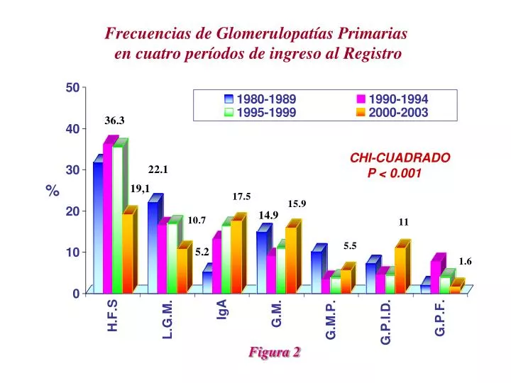
Frecuencias de Glomerulopatías Primarias en cuatro períodos de ingreso al Registro CHI-CUADRADO P < 0.001 Figura 2
Beaufils et al 1978 con SN 7,99 Beaufils et al 1978 sin SN 1,05 Cameron et al 1978 con SN 11,14 Cameron et al 1978 sin SN 1,63 Korbet et al 1986 con SN 5,80 Korbet et al 1986 sin SN 0,83 Velosa et al 1983 con SN 6,73 Velosa et al 1983 sin SN 1,74 FSGS ESRD rates
February 2004 • Volume 43 • Number 2 EditorialPathologic classification of focal segmental glomerulosclerosis: A working proposalVivette D. D’Agati, MDa Agnes B. Fogo, MDb Jan A. Bruijn, MDc Charles Jennette, MDd
Primary (idiopathic) FSGS Secondary FSGS 1. Familial/genetic A. Mutations in -actinin 4 B. Mutations in podocin C. Mutations in WT-1 D. Mutations in integrin 2.Virus-associated A. HIV-1 (HIV-associated nephropathy) B. Parvovirus B19 3. Drug-induced A. Heroin (heroin nephropathy) B. Interferon- C. Lithium D. Pamidronate 4.Mediated by adaptive structural-functional responses A. Reduced renal mass Oligomeganephronia Unilateral renal agenesis Renal dysplasia Reflux nephropathy Sequela to cortical necrosis Surgical renal ablation Chronic allograft nephropathy Any advanced renal disease with reduction in functioning nephrons B. Initially normal renal mass Hypertension Atheroemboli or other acute vaso-occlusive processes Obesity Cyanotic congenital heart disease Sickle cell anemia Table 1. Etiologic Classification of FSGS
Tabla 2.Glosario de términos Adhesión: Continuidad de la matriz de colágeno entre el ovillo glomerular y la cápsula de Bowman Colapso:Colapso implosivo de la pared capilar con plegamiento de la membrana basal glomerular causando obliteración de la luz capilar sin aumento de la matriz o células intracapilares Confluencia de los podocitos con las células parietales o epiteliales tubulares en la luz tubular o cuello: contacto celular directo de los podocitos con las células epiteliales parietales o las células tubulares epiteliales en la luz tubular o el cuello
Hipercelularidad endocapilar: Oclusión de los capilares glomerulares por un aumento absoluto en el número de las células intraluminales, que pueden incluir células espumosas, células endoteliales, macrófagos, y otros leucocitos, a veces asociada con hialinosis, kariorrexis, y raramente fibrina, frecuentemente produciendo una lesión expansiva Focal: Que compromete algunos pero no todos los glomérulos Global: Que afecta todo el ovillo glomerular Glomerulomegalia: Ovillo (Tuft) glomerular de mayor tamaño que los controles de la misma edad ( con la medida de área glomerular > 1.5 x normal)
Hialino: acumulación de material proteinaceo eosinofílico homogéneo “vidrioso” (smooth and glassy) Hipercelularidad Mesangial: >de 3 células mesangiales rodeadas por matiz mesangial en un segmento glomerular intacto, observado en cortes de 3 µm de espesor Perhiliar: Compromiso segmentario vecino al hilio glomerular Hiperplasia podocitaria:Aumento del número de los podocitos, usualmente con acumulación y formación de multicapas; a diferencia de las semilunas verdaderas, estas lesiones típicamente carecen de eje celular, matriz peri-celular, fibrina extracapilar o continuidad con las células epiteliales parietales
Hipertrofia podocitaria: aumento del tamaño de los podocitos con o sin gotas de reabsorción proteica intracitoplasmática, vacuolas, agrandamiento y vesiculización nuclear y nucléolos prominentes Esclerosis: Aumento de la matriz glomerular extracelular con obliteración de la luz capilar glomerular Segmentaria: compromiso del ovillo glomerular < 100% con algunos capilares glomerulares evidentes Dominio TIP: porción externa del ovillo glomerular (25%) cercana al origen del túbulo proximal (el polo tubular debe ser identificado) Ovillo (Tuft): El flóculo glomerular, no incluye la cápsula ni el espacio de Bowman
Propuesta • Clasificación morfológica • Para profundizar en etiología debe realizarse inmunofluorescencia y M.Electrónica • Inicialmente debe descartarse HFS secundaria a otras glomerulopatías • Base para evaluar posibles evoluciones diferentes
CLASIFICACIÓN • NOS (clásica) • Perhiliar • Celular • Lesión Tip • Colapsante
FSGS (NOS)(Not Otherwise Specified)- clásica- • Criterios de Inclusión: • Por lo menos 1 glomérulo con aumento segmentario de la matriz obliterando la luz capilar • Puede existir colapso segmentario de la pared capilar glomerular sin hiperplasia podocitaria por encima • Criterios de exclusión • Variante Perhiliar • Variante Celular • Variante Tip • Variante Colapsante
(A) FSGS (NOS). A low-power view shows segmental lesions ofsclerosis in all 4 glomeruli pictured. Lesions of sclerosis are characterized by increased matrix, causing obliteration ofthe capillary lumina. Distribution of lesions within the tuft is variable, affecting both peripheral and perihilar segments. Insome glomeruli, the location of the sclerosis cannot be ascertained because of the plane of section. (JMS; originalmagnification 100.) (B) FSGS (NOS). High-power view of a glomerulus shows a discrete segmental lesion of sclerosiswith wrinkling and collapse of glomerular basement membranes (stained blue) and hyaline deposits (stained red).Overlying the wrinkled capillaries, there is detachment of podocytes, which form a “cap,” with intervening deposition oflooser neomembrane that is weakly trichrome-blue positive. Although the capped podocytes appear hypertrophied,there is no podocyte hyperplasia. (Masson trichrome; original magnification400.)
(C) FSGS (NOS). There is segmentalobliteration of the glomerular tuft by increased matrix and hyaline material. Sclerosed segments form adhesions toBowman’s capsule. There is no obvious podocyte hypertrophy or hyperplasia. The location of the sclerotic lesion withinthe glomerular tuft cannot be determined because neither the tubular nor the vascular pole are represented in the planeof section. (PAS; original magnification250.)
FSGS Perhiliar • Criterios de inclusión • Por lo menos 1 glomérulo con hialinosis perhiliar con o sin esclerosis • >50% de los glomérulos con lesiones segmentarias deben tener esclerosis o hialinosis perhiliar • Criterios de exclusión • Variante Celular • Variante Tip • Variante Colapsante
(D) FSGS perihilar variant. On low-power examination, 1 of the 3 glomerulipictured contains a discrete lesion of segmental sclerosis affecting the vascular pole region. The lesion shows bothincreased matrix (sclerosis) and hyalinosis. There is adhesion of the sclerotic segment to Bowman’s capsule in thevascular pole region. Perihilar lesions were identified in more than 50% of glomeruli with segmental sclerosis. Both theglomerulus with segmental sclerosis and the 2 nonsclerotic glomeruli are hypertrophied in this patient with morbidobesity. (PAS; original magnification 100.) (E) FSGS perihilar variant. High-power view of the perihilar lesion in (D)shows both the increased matrix (sclerosis) and glassy hyalinosis deposited in the vascular pole segment of the tuft,identified by the incoming arteriole and macula densa. There is no obvious podocyte hypertrophy or hyperplasia. (PAS;original magnification 250.)
(F) FSGS perihilar variant. A very early lesion illustrates the mild increase in matrix andhyaline surrounding the glomerular hilus. There also is hyalinosis of the adjacent preglomerular arteriole. Theglomerulus is hypertrophied in this patient with a solitary functioning kidney. (PAS; original magnification250.)
FSGS Celular • Criterios de Inclusión • Por lo menos 1 glomérulo con hipercelularidad segmentaria endocapilar ocluyendo la luz con o sin células espumosas o kariorrexis, comprometiendo por lo menos 25% del ovillo • Habitualmente existe hipertrofia e hiperplasia podocitaria pero no es un hecho requerido para el diagnóstico • Criterios de exclusión • Variante Tip • Variante Colapsante
FSGS cellular variant. (A) The defining glomerulus in this patient with primary FSGS shows segmentalendocapillary hypercellularity. Involved segments are engorged with endocapillary cells, including some infiltratingmononuclear leukocytes. (H&E; original magnification 250.) (B) In the cellular variant, silver stain showsexpansion of the involved segments by endocapillary cells (not matrix), including foam cells and some karyorrhecticleukocytes. No glomerular capillary wall collapse is seen. There is hypertrophy and hyperplasia of the overlyingpodocytes. (JMS; original magnification 400.)
(C) This example of the cellular lesion shows numerous endocapillaryleukocytes, mimicking a segmental endocapillary proliferativeglomerulonephritis. There is hypertrophy andhyperplasia of overlying podocytes. Adjacent glomerular segments have mild mesangial hypercellularity. (H&E;original magnification 400.) (D) High-power view of a cellular lesion shows karyorrhectic nuclear debris, admixedwith foam cells and hyaline material. Overlying podocytes are hyperplastic. (H&E; original magnification 600.)
FSGS Tip • Criterios de inclusión • Por lo menos 1 lesión segmentaria que compromete el dominio tubular (Tip) (25% externo del tuft cercano al origen del túbulo proximal) • El polo vascular debe estar identificado en la lesión • La lesión debe tener o una adhesión o confluencia de podocitos con células parietales o tubulares en la luz o el cuello tubular • La lesión puede ser celular (<50% ovillo) o esclerosante (<25% ovillo) • Criterios de exclusión • Variante colapsante • Cualquier lesión perhiliar
FSGS tip variant. (A) Low-power view shows a segmental lesion involving the tip domain at the origin ofthe tubular pole. The vascular pole is not visible in this plane of section. (PAS; original magnification 250.) (B)High-power view of the lesion in A shows the involved segment to contain endocapillary foam cells and form anadhesion to Bowman’s capsule at the mouth of the proximal tubule. There is capping of the overlying podocytes,which become confluent with the adjacent tubular epithelium. (PAS; original magnification 600.)
(C) Low-powerview shows the portion of the glomerular tuft involving the tip domain to be expanded by a segmental lesion withcellular features located at the tubular pole. (PAS; original magnification250.) (D) High-power view of the tip lesionin C shows endocapillary foam cells admixed with a few leukocytes. There is a small adhesion of the tuft toBowman’s capsule at the tubular pole. Overlying podocytes appear confluent with the adjacent tubular epithelialcells. (PAS; original magnification 600.)
(E) Opposite the vascular pole, the origin of the proximal tubule is visible.In this area, there is an early tip lesion with inframembranous hyalinosis, a few endocapillary foam cells, andconfluence of swollen podocytes with the tubular epithelium. (PAS; original magnification 250.) (F) In thisglomerulus, the tip lesion appears to herniate into the tubular lumen and forms a capsular adhesion. (PAS; originalmagnification 250.)
FSGF Colapsante • Criterios de inclusión • Por lo menos 1 glomérulo con colapso segmentario o global con hipertrofia e hiperplasia podocitaria por encima • Criterios de exclusión • No
FSGS collapsing variant. (A) A low-power view shows 4 glomeruli, all of which have global collapse of thetuft and podocyte hypertrophy and hyperplasia. There are associated tubular degenerative changes. (JMS; originalmagnification 100.) (B) On high-power view, the lesion of collapsing sclerosis shows global occlusion of capillarylumina by implosive collapse of glomerular basement membranes. There is no appreciable increase in intracapillarycells or matrix. Overlying podocytes form a cellular corona over the collapsed tuft. Some of the enlarged podocytesappear binucleated and have lost their cohesion to the tuft. (JMS; original magnification 400.)
(C) An example ofglobal collapse of the glomerular tuft with marked overlying podocyte hyperplasia, forming a pseudocrescent thatfills the urinary space. An apoptotic podocyte has detached from the tuft and is passing into the tubular pole. (JMS;original magnification 400.) (D) Hyperplastic podocytes contain numerous intracytoplasmic protein resorptiondroplets, which appear red with trichrome stain. The collapsed tuft, shown in blue, has no patent capillary lumina.(Masson trichrome; original magnification 400.)
(E) Hyperplastic podocytes form several layers of cells over thecollapsed tuft. Some of the swollen podocytes contain intracytoplasmic vacuoles. Podocyte nuclei have a vesicularchromatin pattern with prominent nucleoli. (JMS; original magnification 400.) (F) A segmental lesion of collapsingsclerosis spares more than half the tuft. Collapsed capillaries have prominent overlying podocyte hypertrophy andhyperplasia, with abundant trichrome-red intracytoplasmic protein resorption droplets. (Masson trichrome; originalmagnification 400.) (G) By electron microscopy, the collapsed loop has folded glomerular basement membranes,with complete effacement of foot processes. Overlying podocytes are detached from the glomerular basementmembrane with intervening layering of neomembrane material. (Original magnification 2,500.)