Download
1 / 75
770 likes | 1.11k Vues
Anatomy of Phonation. Ch. 4. Spoken Communication. Voiceless phonemes or speech sounds- produced without the use of the vocal folds /s/, /f/ Voiced sounds are produced by action of the vocal folds /z/, /v/ Phonation is voicing, the product of vibrating vocal folds

E N D
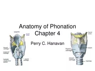
Anatomy of Phonation Ch. 4
Spoken Communication • Voiceless phonemes or speech sounds- produced without the use of the vocal folds /s/, /f/ • Voiced sounds are produced by action of the vocal folds /z/, /v/ • Phonation is voicing, the product of vibrating vocal folds • Respiration is the energy source that permits phonation to occur
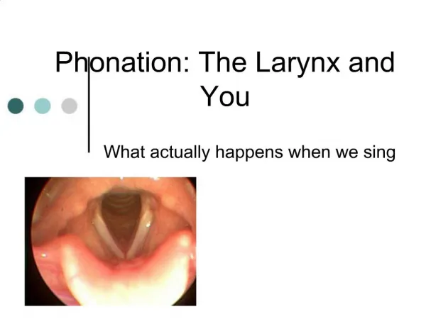
Vocal Folds • Made up of five layers of tissue with the deepest layer being muscle • Space between the vocal folds is glottis • Area below the vocal folds is subglottal region • Alternately produce /a/ and /h/- you should feel the vocal folds vibrate and then stop
Functions of the Larynx • Phonation • Sphincter-vocal folds are capable of a strong and rapid clamping of the airway to keep foreign objects out • Hold your breath • Lifting heavy objects/Childbirth- “fix” or anchor your larynx which provides muscles of upper body a solid framework
Framework of Larynx • A musculocartilaginous structure • Comprised of three unpaired and three paired cartilages bound by ligaments and lined with mucous membrane • Oddly shaped box that sits atop the last ring of the trachea • Adjacent to cervical vertebrae 4-6 in the adult • Average length is 44mm- males, 36 mm- females
Structure of Larynx • Cricoid cartilage • Complete ring resting atop the trachea • Most inferior of the laryngeal cartilages • Side-view looks like a signet ring with the back arching up • Articulates with the thyroid cartilage at the cricothroid joint
Structure of Larynx • Thyroid cartilage • Largest of the cartilages • Articulates with the cricoid cartilage below • Paired processes allow it to rock forward and backward • Arytenoid cartilages • Ride on the high-backed upper surface of the cricoid cartilage • Form the posterior point of attachment for the vocal folds
Structure of Larynx • Corniculate cartilages • Ride on the superior surface of each arytenoid cartilage • Prominent landmarks in the aryepiglottic folds • Cuneiform cartilage resides in the aryepiglottic folds to provide a degree of rigidity • Hyoid bone- articulates to the thyroid cartilage by means of a pair of superior processess • Epiglottis- Medial to hyoid bone and thyroid cartilage, a leaf-like cartilage
Laryngectomy • Surgical removal of larynx • Lose voicing source for speech • Oral cavity is sealed off from the trachea • Breathe through a tracheostoma-opening placed in the trachea • Difficulty with expectoration and coughing • Cannot swim • High risk for foreign objects entering the airway • Flavor of foods is greatly reduced because you do not breathe through the nose • Extreme dryness of oral tissues
Inner larynx • Cavity is a constricted tube • Smooth surface • Sheets and cords of ligaments and membrane • Lines the entire structure with a wet, smooth mucous membrane
Extrinsic ligaments • Thyrohyoid membrane- stretches between the greater cornu of the hyoid and lateral thyroid • Lateral thyrohyoid ligament-superior cornu of the thyroid to posterior tip of the greater cornu hyoid • Median thyrohyoid ligament- from corpus hyoid to upper border of the anterior thyroid • Together- these three connect the larynx and the hyoid bone
Extrinsic ligaments • Hyoepiglottic ligament/Thyroepiglottic ligament- attach the epiglottis to hyoid and inner thyroid cartilage, just below the notch • Cricotracheal ligament-attaches the trachea to the larynx • Lateral and median glossoepiglottic ligaments attach the epiglottis to the tongue • Overlay of the mucous membrane on these ligaments produce the valleculae between the tongue and epiglottis
Intrinsic ligaments • Connect the cartilages of the larynx and form the support structure for the cavity of the larynx and vocal folds • Quandrangular membranes • Layer of connective tissue running from the arytenoids to the epiglottis and thyroid cartilage • Form false vocal folds • Originate at inner thyroid angle and sides of epiglottis and form an upper cone that narros and terminates at the arytenoid and corniculate cartilages
Intrinsic ligaments • Aryepiglottic muscles • From the side of the epiglottis to the arytenoid • Form the upper margin of the quadrangular membrane • Form the aryepiglottic folds • Folds are simply the ridges marking the highest elevation of these membranes • Pyriform sinus is the space between the aryepiglottic fold and the thyroid cartilage
Vocal Fold Structure • Composed of five layers of tissue p. 174-177 • Most superficial layer- protective layer of squamos epithelium, gives them a glistening white appearance, keeps them moist by assisting in fluid retention • Second layer- superficial lamina propria (SLP)- elastin fibers, allows them to be extensively stretched, cushions the vocal folds • Third layer- Intermediate lamina propria (ILP)- elastin fibers running in an A-P direction, cross layered with the SLP, combination of these two layers provide elasticity and strength
Vocal Fold Structure • Fourth layer- Deep lamina propria (DLP)- made of collagen fibers that prohibit extension. • ILP and DLP combine to make up the vocal ligament • Fifth layer-thyroarytenoid muscle- makes up the bulk of the vocal fold, muscle runs A-P, active element of the vocal folds • Mucosal lining- combination of the epithelial lining and first layer • Vocal ligament- second and third layers which gives a degree of stiffness and support • Body- fourth layer and muscle
Cavities of Larynx • Aditus- entry to the larynx from the pharynx above • Vestibule- space between the aditus and the ventricular folds (false vocal folds) • False vocal folds- not used for phonation except in rare cases • Vestibule is wide at the aditus and narrows • Lateral walls are comprised of the aryepiglottic folds • Posterior walls are made up of the membrane covering the arytenoid cartilages • Rima vestibuli- space between the false vocal folds
Cavities of Larynx • Laryngeal ventricle- middle space of the larynx • Lies between the margins of the false vocal folds ad true vocal folds • Contains the laryngeal saccule which has more than 60 mucous glands that secrete lubricating mucous into the laryngeal cavity • Glottis- space between vocal folds • Variable sphincter that permits voicing • Length at rest is 20mm in adults from the anterior commissure to the posterior commisure
Cricoid Cartilage • Unpaired cartilage • Most inferior of the cartilages • Higher in back than in front • Posterior quadrate lamina- provides point of articulation for arytenoid cartilages • Lateral surface- point of articulation for inferior horns of thyroid cartilage • Cricothyroid joint- diathrodial, pivoting joint permits rotation of the two structures • Would fit loosely on your little finger
Thyroid Cartilage • Unpaired cartilage • Largest • Prominent anterior surface made of two places- thyroid laminae • Joined at the midline- thyroid angle • Superior most point is the thyroid notch (adam’s apple)
Thyroid Cartilage • Vocal folds attach to the thyroid just behind the thyroid notch • Posterior aspect is open- two prominent sets of cornu or horns • Inferior cornua project downward to articulate with the cricoid cartilage • Superior corner project superiorly to articulate with the hyoid • P. 181
Arytenoid Cartilage • Paired cartilage • Among the most important • Superior posterolateral surface of cricoid cartilage • Mechanical structure that permits onset and offset of voicing • Formed like a pyramid • Each cartilage has two processes and four surfaces
Arytenoid Cartilage • Apex- superior portion • Corniculate cartilage sits on the superior surface • Base- inferior surface • Concave surface- point of articulation with the cricoid cartilage • Two processes • Vocal processes project anteriorly toward thyroid notch, posterior portion of the vocal folds attach • Muscular process forms the lateral outcropping of the arytenoid pyramid- point of attachment for muscles that adduct/abduct the vocal folds
Epiglottis • Unpaired cartilage • Leaf-like structure arises from the inner surface of the thyroid cartilage just below the notch • Attached by the thyroepiglottic ligament • Sides are joined with the arytenoid artilages via the aryepiglottic folds • Projects upward beyond the larynx and above the hyoid bone
Epiglottis • Attached to the root of the tongue by glosso-epiglottic fold and lateral glosso-epiglotticligamets • This juncture produces the valleculae • Pyriform sinuses- lateral recesses • Attached to the hyoid bone via the hyoepiglottic ligament • Surface is covered with a mucous membrane lining • Beneath this lining there are branches of the internal laryngeal nerve of the X vagus that conducts sensory information from the larynx
Cuneiform cartilages • Small cartilages embedded within the aryepiglottic folds • Situated above and anterior to the corniculate cartilages • Provide support for the membranous laryngeal covering
Hyoid Bone • Provides the union between the tongue and the laryngeal structure • Unpaired small bone • Articulates loosely with the superior cornu of the thyroid cartilage • U-shaped, being open in the poseterior side • Corpus or Body- forms the front of the bone, point of attachment for six muscles • Greater cornu- lateral surface of the corpus projecting posteriorly • Lesser cornu-found at the junction of the corpus and greater cornu
Movement of the Cartilages • Cricothyroid and Cricoarytenoid joints are the only functionally mobile points of the larynx • Cricothyroid joint- junction of cricoid cartilage and inferior cornu of the thyroid cartilage • Diarthrodial (synovial) joint that permit the cricoid and thyroid to rotate and glide • Rotation permits the thyroid cartilage to rock down in front • Permits the thyroid cartilage to glide forward and backward slightly • Provides the major adjustment for change in vocal pitch
Movement of the Cartilages • Cricyarytenoid joint • Formed between the cricoid and arytenoid cartilages • Synovial joints permit rocking, gliding and minimal rotation • Rocking action permits two vocal processes toward each other permitting the vocal folds to approximate • Arytenoids glide on the long axis which changes vocal fold length • Arytenoids rotate upon a vertical axis which permits abduction
Intrinsic Laryngeal Muscles • Have both origin and insertion on laryngeal cartilages • Make fine adjustments to the vocal mechanism • Assume responsibility for opening, closing, tensing, and relaxing the vocal folds
Intrinsic Laryngeal Muscles • Lateral Cricoarytenoid Muscle • Attaches to the cricoid and the muscular process of arytenoid • Muscular process will be drawn forward • Rocks the arytenoid inward and downward • Adduction of vocal folds • Lengthens the vocal folds • Innervated by X vagus nerve- arises from medulla oblongata
Intrinsic Laryngeal Muscles • Transverse Arytenoid Muscle • Runs between the two arytenoid cartilages on the posterior surface • Pulls the two arytenoids closer together • Approximates the vocal folds • Increased medial compression which is increased force of adduction • Vital element in vocal intensity change • Innervation by the inferior branch of the Recurrent laryngeal nerve of the X vagus
Intrinsic Laryngeal Muscles • Oblique Arytenoid Muscles • Immediately superficial to the transverse arytenoid muscles • Perform a similar function • Paired muscles • Originate at the posterior base of the muscular process and course obliquely up to the apex of the opposite arytenoid • Form an “X” • Pull the apex medially • Promotes adduction • Enforces medial compression • Rocks arytenoid and vocal folds down and in • Aids in pulling the epiglottis to cover the opening to the larynx • Innervation- Recurrent laryngeal nerve of the X Vagus
Intrinsic Laryngeal Muscles • Posterior Cricoarytenoid muscle • Sole abductor of the vocal folds • Originates from the posterior cricoid lamina • Project up to insert into the posterior aspect of the muscular process of the arytenoid cartilage • Pulls muscular process posteriorly • Rocks the arytenoid cartilage • Abducts the vocal folds
Intrinsic Laryngeal Muscles • Cricothyroid Muscle- composed of two heads • Pars Recta- originates on the anterior surface of cricoid cartilage and courses up to the lower surface of the thyroid lamina • Pars oblique- arises from the lateral cricoid cartilage to the point of juncture between the thyroid laminae and inferior horns • Both tense the vocal folds • Together they are the major contributors for pitch change • Innervated by the Superior Laryngeal Nerve of the X Vagus
Intrinsic Laryngeal Muscles • Pars Recta • Rocks the thyroid cartilage downward • Brings thyroid and cricoid closer together in front • Makes the posterior cricoid more distant from the thyroid • Vocal folds are stretched • Pars Oblique • Thyroid slides forward • Tense the vocal folds
Intrinsic Laryngeal Muscles • Thyrovocalis Muscle (abbreviated as vocalis) • Medial muscle of the vocal folds • Originates from the inner surface of the thyroid cartilage • Inserts into the lateral surface of the arytenoid vocal process • Contraction draws the thyroid and cricoid cartilages farther apart in front • Glottal tensor- tenses the vocal folds • Innervated by the Recurrent Laryngeal Nerve of the X Vagus
Intrinsic Laryngeal Muscles • Thyromuscularis Muscle • Paired muscle • Immediate lateral to each vocalis muscle • Originates on the inner surface of the thyroid cartilage near the notch • Inserts into the arytenoid cartilage at the base • Laryngeal relaxer • Relaxes the vocal folds • Innervated by the Recurrent Laryngeal Nerve
Intrinsic Laryngeal Muscles • Movement of the vocal folds into and out of approximation is achieved by the coordinated effort of many of the intrinsic muscles of the larynx • Vocal folds are also lengthened which increases the tension on them • Changing pitch is reflected by a change in mass or tension