Download
1 / 34
360 likes | 369 Vues
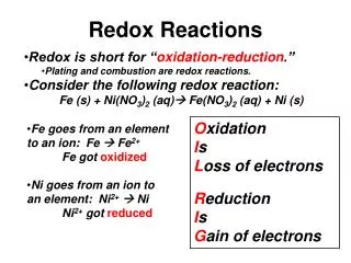
Emory Clinical Biomarkers Laboratory. Measuring the poise of thiol/disulfide redox in vivo. Dean P. Jones, Ph.D. Department of Medicine/Division of Pulmonary, Allergy and Critical Care Medicine Emory University, Atlanta. GSH. GSSG. [Ox]_. RT n F. E. = E. + * ln. h. o.

E N D
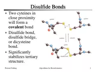
Emory Clinical Biomarkers Laboratory Measuring the poise of thiol/disulfide redox in vivo Dean P. Jones, Ph.D. Department of Medicine/Division of Pulmonary, Allergy and Critical Care Medicine Emory University, Atlanta
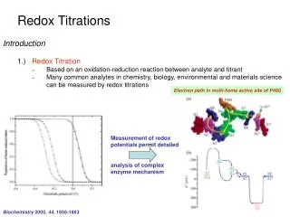
GSH GSSG [Ox]_ RT nF E = E + * ln h o [Red] The redox state of GSH/GSSGprovides a measure of the balance of prooxidants and antioxidants Redox states of different couples can be compared by expression as redox potentials Low molecular weight thiols and disulfides are measured by HPLC Jones Meth Enzymol 2002
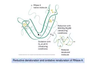
Reversible oxidation of thiols alters protein structure and function Reduced Trx1 Oxidized Trx1 Active site Watson et al. 2003
Active site ASK-1 docking regulatory site Dimerization site S-Nitrosylation site S-Glutathiylation site, S-Alkylation site Redox "OFF" switch All cysteines in Trx1 are important in Trx1 function
Trx1R Trx1O Trx2R Trx2O Protein thiol/disulfide redox states are measured by Redox Western blot analysis Native gel separation by charge following treatment with IAA Trx1-Ox2 Trx1-Ox1 Trx1-Red Nuclei Trx1-Ox2 Trx1-Ox1 Trx1-Red Cytoplasm 0 2 10 30 60 120 Time (min) after H2O2 Watson, Jones FEBS Lett 2003 Watson et al, JBC 2003 SDS-PAGE separation by mass following treatment with AMS Cytoplasm Mitochondria 0 50 200 300 400 µM tBH Chen et al FEBS Lett 2006
Quantification of thiol/disulfide redox in biologic systems has provided 3 general conclusions 1. At the cellular level, GSH redox becomes oxidized as cells progress through the life cycle, and cells regulate extracellular thiol/disulfide redox state 2. At the systemic level, plasma GSH redox becomes oxidized with oxidative stress and is oxidized in association with aging and chronic disease 3. In cells and plasma, GSH redox is NOT equilibrated with thioredoxin or Cys/CySS, providing the basis to consider discrete redox circuits for redox signaling and control
Redox of GSH/GSSG becomes progressively oxidized in the life cycle of cells -(SH)2:-SS- Proliferation 100:1 - - 250 250 , mV) h 10:1 Differentiation Redox State (E Redox State (E 1:1 - - 200 200 Apoptosis 1:10 - - 150 150 Kirlin et al, FRBM 1999; Nkabyo et al, Am J Physiol 2002
Eh, GSH/GSSG Mean=-130.9 StDev=22.9 -120 +200 mM Cysteine -100 -80 -60 -40 +100 mM Cystine Eh, Cys/CySS -20 Mean=-72.4 StDev=12.8 Time (h) 4 8 12 16 20 24 0 Extracellular Cys/CySS pool in culture is regulated to a value very similar to that in human plasma HT29 cells Plasma, 740 subjects Frequency Extracellular Eh (Cys/CySS) (mV) Eh (mV) Jonas et al, FRBM 2002 Go and Jones, Circulation 2005
Interorgan GSH/Cysteine balance Tissues Plasma Major pool Most reduced -220 mV -138 mV GSH/GSSG GSH/GSSG Cys/CySS -150mV Cys/CySS -80 mV Major pool Most oxidized
Quantification of thiol/disulfide redox in biologic systems has provided 3 conclusions 1. At the cellular level: Cells regulate extracellular thiol/disulfide redox state. Cellular GSH redox becomes oxidized as cells progress through the life cycle 2. At the systemic level: Plasma GSH redox becomes oxidized with oxidative stress. Plasma redox is oxidized with aging, nutritional deficiency, toxicity and chronic disease 3. Relationship of redox couples: GSH redox is NOT equilibrated with thioredoxin or Cys/CySS. This provides the basis to consider discrete redox circuits for redox signaling and control
Eh, GSH/GSSG Mean=-130.9 StDev=22.9 Eh, Cys/CySS Mean=-72.4 StDev=12.8 4 8 12 16 20 24 0 Many people have redox states more oxidized than young healthy individuals HT29 cells Plasma, 740 subjects Young healthy in RED -120 +200 mM Cysteine Reduced -100 -80 Frequency Oxidized Reduced Extracellular Eh (Cys/CySS) (mV) -60 Oxidized -40 +100 mM Cystine -20 Eh (mV) Time (h) Jonas et al, FRBM 2002 Go and Jones, Circulation 2005
- - 130 130 p = 0.001, effect of time p = 0.001, effect of time - - 120 120 (mV) (mV) chemo chemo chemo chemo h h E E - - - - 110 110 - - Day 10 Day 10 Day 14 Day 14 Day 3 Day 3 Day 7 Day 7 Post Post Pre Pre - - 100 Plasma redox provides a useful measure of oxidative stress in humans GSH & Cys redox oxidized with age Cys redox oxidized with smoking - - 90 90 - - 80 80 * * Eh Cys (mV) - - 70 70 - - 60 60 N = 66 66 64 64 53 53 Never Never Prior Prior Current Current Smoking Status Jones, FRBM 2002 Moriarty, FRBM 2004 GSH redox is oxidized with chemotherapy Antioxidants decrease Cys oxidation with age - - 140 140 Mean age = 71.7 Mean age = 71.7 - - 120 120 +Vit C, E, b-car GSH (mV) Control P = 0.002 for effect of time P = 0.002 for effect of time Mean age = 76.3 Mean age = 76.3 - - 100 100 h E 70 70 72 72 74 74 76 76 78 78 Age (y) Jonas, Am J Clin Nutr 2000 Moriarty-Craige, Am J Ophthalmol 2005
GSH/GSSG is oxidized in T2 Diabetes -135 * * ** GSH/GSSG Eh (mV) -110 Type 2 Diabetes <60 >60 Controls Controls Samiec et al, FRBM 1998 Plasma redox is oxidized in association with disease and disease risk Eh GSH/GSSG predicts IMT 0.67 p value 0.009 0.66 0.61 0.62 Carotid IMT (mm) 0.59 0.58 0.54 < -130 mV -120 to -130 mV > -120 mV Eh GSH/GSSG Ashfaq et al, Am Coll Cardiol 2006
Health Pathophysiologic correlation -80 mV Low antioxidants, low dietary cysteine (-140 mV) -62 mV (-122 mV) Aging Alcohol abuse -50 mV (-110 mV) Type 2 Diabetes Cigarette Smoking Increased Carotid Intima Media Thickness Reversible myocardial perfusion defects Chemotherapy/BMT -20 mV (-80 mV) Lung transplantation Cys/CySS Redox (GSH/GSSG Redox) Jones, Antiox Redox Signal, 2006
Quantification of thiol/disulfide redox in biologic systems has provided 3 general conclusions 1. At the cellular level, GSH redox becomes oxidized as cells progress through the life cycle, and cells regulate extracellular thiol/disulfide redox state 2. At the systemic level, plasma GSH redox becomes oxidized with oxidative stress and is oxidized in association with aging and chronic disease 3. In cells and plasma, GSH redox is NOT equilibrated with thioredoxin or Cys/CySS, providing the basis to consider discrete redox circuits for redox signaling and control
- - 300 300 Trx Proliferation Differentiation Apoptosis , mV) - - 250 250 h Proliferation Redox State (E Redox State (E GSH - - 200 200 Differentiation Apoptosis Proliferation Differentiation Cys - - 150 150 GSH, Trx and Cys redox systems are not in redox equilibrium in cells Jones et al FASEB J 2004
-300 -250 -200 -150 GSH/GSSG, Trx and Cys/CySS provide independent nodes for redox signaling and control NADPH TR1 GR Trx 3 GSH/GSSG (proliferation) 6a 1/Prx Eh (mV) GSH/GSSG (differentiation) 2 6b GSH/GSSG (apoptosis) Grx Cys/CySS 4 5/GPx SO H2O2 TO H2O2 O2 O2 Cellular Extracellular Jones et al, FASEB J 2004
Redox-dependent systems are differentially controlled by GSH, Trx1 and Cys redox couples Trx/TrxSS ASK-1 Apoptosis KEAP-1 Nrf-2 translocation to nucleus GSH/GSSG GSH/GSSG Nrf-2 DNA binding EGFR MAPK activation Cys/CySS Trx/TrxSS Cys/CySS Protein synthesis Protein S-thiylation
Compartmentation of thiol/disulfide redox state Plasma/Interstitial Cytoplasmic GSH/GSSG Trx1(-SH)2/SS Cys/CySS GSH/GSSG Cys/CySS Mitochondrial Trx2(-SH)2/SS Nuclear GSH/GSSG Trx(-SH)2/SS GSH/GSSG Endoplasmic Reticulum GSH/GSSG PDI Hansen et al, Annu Rev Pharm Tox, 2006
Trx2 is preferentially oxidized by TNF Thioredoxin-2 Thioredoxin-1 H2O2 (mM) H2O2 (mM) TNF (ng/ml) TNF (ng/ml) 0 5 10 20 40 1 0 5 10 20 40 1 -300 -380 -360 -280 -340 Redox Potential (Eh) Redox Potential (Eh) -320 -260 -300 -280 -240 0 5 10 20 40 H2O2 0 5 10 20 40 H2O2 TNF (ng/ml) TNF (ng/ml) J. Hansen
Mitochondrial redox circuits Metabolic Redox Circuits (high flux) Redox Signaling and Control Circuits (low flux) Metabolic substrates NADPH Eh NADPH GR TR2 -400 ASK1 Pyr NADH Grx2 Mal PrSSG Trx2 GSH -200 MPT Succinate 0 GPx Prx3 CoQ O2- +200 O2- MnSOD Cyt c H2O2 +400 O2 O2 +600 Regulatory Signal DP Jones, Chem-Biol Interact 2006
Summary: Trx2 in Mitochondrial Compartment 1. Mitochondrial Trx2 has a more reduced redox state than cytoplasmic or nuclear Trx1 or cellular GSH 2. Mitochondrial Trx2 is more susceptible to oxidation than the cytoplasmic Trx1 3. Redox western blot analysis of mitochondrial Trx2 provides a useful approach to measure mitochondrial oxidative stress
GSH is difficult to measure in nuclei Cotgreave, 2003 Bellomo, 1992 Voehringer, 1998
Translocation of Trx from the cytoplasm to the nucleus Hirota et al, J Biol Chem (1999) 274:27891
-350 -300 Eh (mV) -250 Trx/TrxSS -200 GSH/GSSG -150 0 10 20 30 40 50 60 Time (min) High levels of oxidants are not selective between GSH and Trx1 Time courses of GSH and Trx1 oxidation are similarTrx-1 is somewhat more resistantTrx-1 recovers somewhat more rapidly +1 mM H2O2 Trx-Ox2 Trx-Ox1 Trx-Red Nuclei Trx-Ox2 Trx-Ox1 Trx-Red Cytoplasm 0 2 10 30 60 120 Time (min) Watson, Jones (2003) FEBS Lett 543:144
Cytosolic Trx1 Nuclear Trx1 -285 -285 -275 -275 Nuclear Trx1 Eh (mV) Cytoplasmic Trx1Eh (mV) -265 -265 -255 -255 0 10 20 30 0 10 20 30 Time (min) Time (min) -365 Mitochondrial Trx Cellular GSH -285 -355 -275 Trx2 Eh (mV) GSH/GSSG Eh (mV) -345 -265 -335 -255 0 10 20 30 0 10 20 30 Time (min) Time (min) Physiologic oxidation in response to EGF is specific to cytosolic Trx-1 P. Halvey et al, Biochem J 2005
Trx1 and PrSH/PrSSG are more reduced in nuclei Nuclei contain less protein-SH per mg protein than cytoplasm Nuclear Trx1 and PrSH/PrSSG are more resistant to oxidation than cytoplasmic pools
Transcriptional activation by Nrf2 Cytoplasm Nrf-2 Keap-1 Nucleus Nrf-2 Nrf-2 Maf Keap-1 Nrf-2 Transcription ARE Maf ARE
200 160 120 80 40 0 Empty TRX1 TRX1 +TBHQ GSH controls cytoplasmic activation of Nrf2 translocation to nucleus Cytoplasm Nrf-2 Keap-1 Nucleus ↓GSH ↑GSH Nrf-2 Nrf-2 Maf Keap-1 Nrf-2 Transcription ARE Maf ARE 300 250 200 Nuclear Nrf-2 (% Control) 150 100 50 0 Control BSO NAC J. Hansen et al, Tox Sci 2004
200 160 300 250 120 200 150 80 100 % Control (Luc/B-gal) 40 50 0 0 Empty Trx-1 Empty TRX1 TRX1 +TBHQ NLS-Trx-1 C35S Trx-1 C35S NLS-Trx-1 GSH and Trx control different steps in transcriptional activation by Nrf2 Cytoplasm Nrf-2 Keap-1 Nucleus Nrf-2 ↓GSH ↑GSH Trx1(SH)2 Nrf-2 Trx1(SS) Maf Nrf-2 Keap-1 Nrf-2 Transcription ARE Maf ARE 300 250 200 Nuclear Nrf-2 (% Control) 150 100 50 0 Control BSO NAC J. Hansen et al, Tox Sci 2004
Cytoplasmic activation of Nrf2 is dependent upon GSH/GSSG Nuclear activity of Nrf2 is dependent upon Trx1
IkB IkB p50 p65 p50 p65 p50 p65 p50 p65 Distinct roles for Trx in the cytoplasm and the nucleus endotoxin cytokines oxidants, etc. Ubiquitination, Degradation PO4 + Trx-(SH)2 cytosol nucleus Ref1 <-- Trx-(SH)2 NF-kB-dependent gene (e.g. TNF)
Plasma/Interstitial GSH/GSSG = -140 Cytoplasmic Cys/CySS = -80 Trx1(-SH)2/SS = -280 GSH/GSSG = -220 to -260 Cys/CySS = -160 Mitochondrial Trx2(-SH)2/SS = -360 Nuclear Trx1(-SH)2/SS = -300 GSH/GSSG = -300 Endoplasmic Reticulum GSH/GSSG = -150 Hansen et al, Annu Rev Pharm Tox, 2006
Summary • Redox signaling and control involves discrete redox circuitry • The mitochondrial compartment is most reduced and most susceptible to oxidation • Nuclei are more reduced than cytoplasm and contain special mechanisms to protect against oxidative stress • Analytic methods are available to elucidate the redox circuitry and compartmentation of oxidative stress