Download
1 / 25
250 likes | 257 Vues
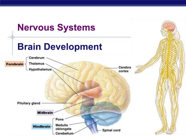
The nervous system: the ear. Emily Torres Ariana Soto Per: 5. Sense of Hearing. The Ear enables us to be able to: - hear sound waves - have sense of equilibrium The Ear is composed of the: - Outer (External) Ear - Middle Ear - Inner (Internal) Ear. Outer (External) Ear.

E N D
The nervous system: the ear Emily Torres Ariana Soto Per: 5
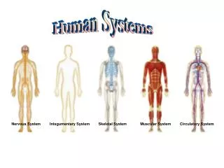
Sense of Hearing • The Ear enables us to be able to: • - hear sound waves • - have sense of equilibrium • The Ear is composed of the: • - Outer (External) Ear • - Middle Ear • - Inner (Internal) Ear
Outer (External) Ear • The Outer Ear consists of these structures: • - Auricle (Pinna) • - External Auditory Canal • - Tympanic membrane • The opening before the temporal bone, there are hairs that guard the canal and skin that contains ceruminous glands. • - Ceruminous glands secrete cerumen (wax) • - wax helps keep foreign particles out
Middle Ear • The middle ear consists of these structures: • - tympanic membrane • - auditory ossicles: • i. Malleus • ii. Incus • iii. Stapes • - oval window • - tensor tympani • - stapedius
Malleus, incus, & stapes • These three bones connect the tympanic membrane and inner (internal) ear by tiny ligaments • Malleus • - vibrates at the same time as tympanic membrane, then causing the incus to vibrate • Incus • - vibrates and sends the vibrations on to the stapes • Stapes • - vibrates and sends a fluid into the inner ear through the oval window
Tensor tympani & stapedius • Tensor tympani • - connected to the wall of the auditory tube • - pulls malleus inward when contracted • Stapedius • - pulls the stapes outward • These involuntary actions are part of the effectors in the tympanic reflex
Tympanic reflex • Occurs in about 1/10th of a second after a loud sound in order to protect the hearing receptors from possible damage from a loud external noise • - also occurs when a person is simply talking or singing
Auditory tube & Ear pressure adjustment • Connects each middle ear to the nasopharynx • Helps provide equal air pressure on each side of the tympanic membrane
Ear Infections • Infection of the middle ear • Most common ear infection is otitis media and is usually a result from: • - common cold • - flu • - allergies • - sinus infection
Inner (Internal) ear • Structures include; • Cochlea • Semicircular canals • Vestibule
cochlea • Fluid filled snail-like structure that contains the receptor organ for hearing • Spiral organ of Corti • Divided into 3 chambers • Contains hair cells • 2 types of fluid • Perilymph • Endolymph
Cochlea Chambers • ScalaVestibuli- • Perilymph (provides cushioning support, similar to interstitial fluid) • Scala Media- • Endolymph • Organ of Corti • Scala Tympani- • Perilymph (provides cushioning support, similar to interstitial fluid)
Organ of corti • Found on basilar membrane in the scala media chamber • Sensory organ of hearing • Sound sensitive hair cells • Pressure change causes hair cells to bend and stimulate a nerve signal which is carried to the brain • This is how sound can be recognized
Semicircular Canal • Body’s balance organ • 3 canals (superior, horizontal, and posterior) filled with endolymph that are each responsible for detecting motion on a different plane • Sense head rotations/movement
Vestibule • Senses movement • Holds 2 sacs of fluid • the ultricle & the saccule • Hair cells • Macula • Known as gravity receptors
Equilibrium • State of balance • Condition in which contending forces are equal • 2 types: • Static • Dynamic
Static equilibrium • Occurs when the body is motionless or moving in a straight line • Maintains stability and posture • Entrance of inner ear is the Vestibule • Vestibule contains 2 sacs of fluid (Ultricle & Saccule) • The maculae of the ultricle and saccule are the gravity receptors that respond to linear acceleration, deceleration, and position in space • Otoliths are calcium carbonate particles found in the fluid • When your head moves, the otolithsbursh against the hair cells that line the utricle & saccule • Sends message to brain
Dynamic equilibrium • Occurs when the body is moving in a rotational or angular direction (spinning) • Detects motion • Sense of balance is interpreted in the Semicircular Canals • Ampullae, the organs of dynamic equilibrium, are located at the base of each semicircular canal • A gelatinous cupula is found in the ampullae • Hair cells found on the cupula respond to movement • When head rotates, endolymph moves and flows into the ampulla and causes the cupula to bend which triggers the hair cells to send messages to the brain
Sound collection & pathway through the ear canal • The Auricle (Pinna) • - collects sound waves in the air, then sendsthem into the auditory canal • The Auditory Canal • - once sound waves enter here and pass to the end of the canal, they alter pressure on the tympanic membrane • The Tympanic Membrane • - moves back and forth due to sound waves,whichthen makes the vibrations
Malleus, incus, & stapes (ossicles) • These three bones connect the tympanic membrane and inner (internal) ear by tiny ligaments • Malleus • - vibrates at the same time as tympanic membrane, then causing the incus to vibrate • Incus • - vibrates and sends the vibrations on to the stapes • Stapes • - vibrates and sends fluid into the inner ear through the oval window
(Continued) The Oval Window - is an opening that transmits pressure waves of sound into the cochlea The Round Window -waves move to the round window and it releases pressure caused by the vibrations of the stapes in the oval window The Cochlea - sound waves make fluid in cochlea move and trigger the hair cells to move as well. The hair cells located on the organ of corti separate the sound waves according to their frequencies
(Continued) The Auditory Nerve - hair cells cause an electrical signal to travel through the auditory nerve and to your brain The Brain - the brain responds to these separate frequencies and makes a complete sound from them
Sources: • "Audition: Hearing, the Ear, and Sound Localization - Boundless Open Textbook." Boundless. N.p., n.d. Web. 30 Apr. 2015. • "How We Hear A Sound?" YouTube. YouTube, n.d. Web. 30 Apr. 2015. • "Ear Ossicles." Ear Ossicles. N.p., n.d. Web. 30 Apr. 2015. • "Hearing and the Cochlea - Anatomy Video: MedlinePlus Medical Encyclopedia." U.S National Library of Medicine. U.S. National Library of Medicine, n.d. Web. 30 Apr. 2015. • "Sense of Balance." Flashcards. N.p., n.d. Web. 30 Apr. 2015. • "The Cochlea - HowStuffWorks." HowStuffWorks. N.p., n.d. Web. 30 Apr. 2015. • "Middle Ear." Middle Ear Anatomy, Function & Diagram. N.p., n.d. Web. 30 Apr. 2015. • "Ear Infection Facts: Causes, Acute Infections, Chronic Infections, and More."WebMD. WebMD, n.d. Web. 30 Apr. 2015. • "Pharyngotympanic Tube." Eustachian Tube Function, Anatomy & Diagram. N.p., n.d. Web. 30 Apr. 2015. • "See Pictures of Ear Infection Causes, Symptoms and Treatment."MedicineNet. N.p., n.d. Web. 30 Apr. 2015. • "Anatomy of the Ear." Anatomy of the Ear. N.p., n.d. Web. 30 Apr. 2015.