Download
1 / 58
580 likes | 706 Vues
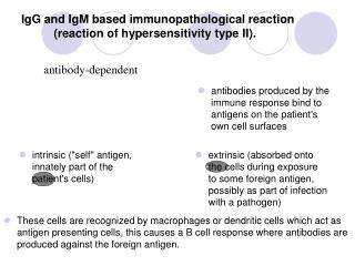
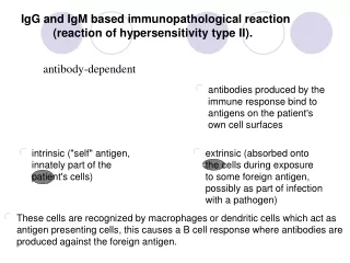
IgG and IgM based immunopathological reaction (reaction of hypersensitivity type II). = antibody-dependent. antibodies produced by the immune response bind to antigens on the patient's own cell surfaces. intrinsic ("self" antigen, innately part of the patient's cells).

E N D
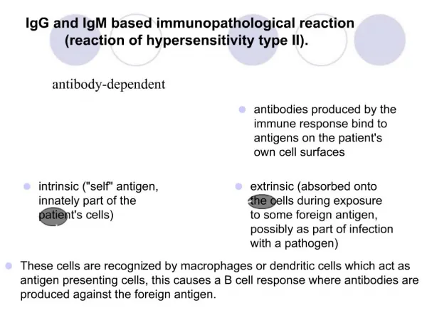
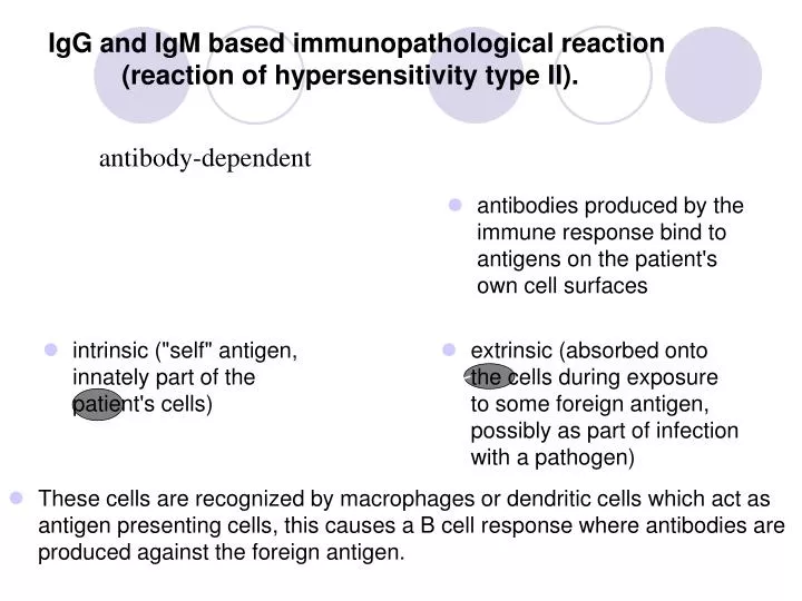
IgG and IgM based immunopathological reaction (reaction of hypersensitivity type II). • = antibody-dependent • antibodies produced by the immune response bind to antigens on the patient's own cell surfaces • intrinsic ("self" antigen, innately part of the patient's cells) • extrinsic (absorbed onto the cells during exposure to some foreign antigen, possibly as part of infection with a pathogen) • These cells are recognized by macrophages or dendritic cells which act as antigen presenting cells, this causes a B cell response where antibodies are produced against the foreign antigen.
IgG and IgM based immunopathological reaction (reaction of hypersensitivity type II) • Autoimmune hemolytic anemia • Goodpasture's syndrome • Autoimmune pernicious anemia • Immune thrombocytopenia • Transfusion reactions • Myasthenia gravis • Rheumatic fever • Acute transplant rejection
Immune complex based immunopathological reaction (reaction of hypersensitivity type III) • occurs when antigens and antibodies are present in roughly equal amounts, causing extensive cross-linking • large immune complexes that cannot be cleared are deposited in vessel walls and induce an inflammatory response • the reaction can take hours, days, or even weeks to develop
Immune complex based immunopathological reaction (reaction of hypersensitivity type III) Some clinical examples: • Rheumatoid arthritis • Immune complex glomerulonephritis • Serum sickness • Subacute bacterial endocarditis • Systemic lupus erythematosus • Farmer's lung (Arthus-type reaction) • Polyarteritis nodosa
PRIMARY IMMUNODEFICIENCY clinical manifestactions examples
IMMUNODEFICIENCY • Primary immunodeficiencies - congenital, genetically defined disorders - onset of symptoms - predominantly at an early age • Secondary immunodeficiencies - caused by chronic infections, irradiation, injuries, immunosupression therapy, surgery, stress - disorders appear at any age
IMMUNIDEFICIENCY • Humoral deficiency disorders = the B cell deficiency disorders – the qualitative or quantitative defects of the B cells, present 70% of IDs • T cell deficiency disorders and the combined B-cell and T-cell deficiency disorders (20%) – group of the qualitative or quantitative defects of the T and B cells • Phagocytic celldisorders– group of the qualitative or quantitative defects of the fagocytic cells (10%) • Complement disorders – caused by the deficiency of the complement components or functions (<1%)
MAJOR CLINICAL FEATURES • Humoral deficiency disorders - manifest as the recurrent bacterial sinopulmonary and gastrointestinal infections - caused by streptococcus, staphylococcus, haemophilus, begin when infants are 5-9 months of age • T cell disorders - manifest as the recurrent bacterial, fungal and viral respiratory and gastrointestinal infection • Complement disorders – are associated with increased incidence of the infections and autoimmune diseases and with edema in the case of hereditary angioedema • Phagocytic celldisorders – characterized by recurrent infections caused by various organisms incluging abscesses, purulent skin infections, granulomatousinflammations
HUMORAL DEFICIENCY DISORDERS • Bruton’s X-linkedhypogamaglobulinemia • CVID - Common Variable ImmunoDeficiency • Selective immunoglobulin A deficiency <0,07 g/l
Bruton’s X-linkedhypogamaglobulinemia • the genetic defect on the X chromosome leads to the defective function of a tyrosine kinase in the B cells • This defect result in a block of the pre-B cells maturation into the B cells with surface IgM • the immunologic findings: < 2% circulating B cells - low serum levels of all classes of immunoglobulins - number and function of T cells are intact - pre-B cells are in the bone marrow • features : begining from 5-9 months of age - manifests as recurrent bacterial sinopulmonary and gastrointestinal infection caused by streptococcus, staphylococcus, haemophilus, meningococcus, salmonella, campylobacter, giardia • Treatment consists of life-long intravenous pooled human gammaglobulin replacement and antibiotics.
Common VariableImmunoDeficiency • the B cell functional disorder characterized by the normal number of the B cells, low levels of IgG and IgA, a poor response to all vaccines and decrease of the T cells (CD4+) number and function • the symptom’s onset between 2ndand 3rddecade • the clinical features: - recurrent respiratory tract infections (pneumonia), cutaneous and gastrointestinal infection - disease is accompanied by occurrence of the granulomas, lymphadenopathy, splenomegaly • Treatment consist of the intramuscular or intravenous gammaglobulin replacement.
Selective deficiency of IgA • level of IgA up to 0,05 g/l, age > 4 years • the most frequent primary ID - stem cell defect - repeated infections of respiratory tract - susceptibility to autoimmune disorders, malignant disorders, allergy - contra-indication of administration of drug with IgA
T cell deficiency disorders • DiGeorge syndrome - the genetic defect on the chromosome 22 leads to disorder of development of 3rdand 4thbranchial pouch with congenital hypoplasia of both the thymus and parathyroid glands - patients suffer from disorder of pre-thymocytes maturation due to absence/hypoplasia of thymus - syndrome CATCH 22: cardiac defects, abnormal facies, thymic hypo/aplasia, cleft palate, hypocalcemia, deletion 22q11.2 - the symptom’s onset soon after the birth – hypocalcemic spasms andmanifestations of congenital heart disease - treatment: symptomatic, transplantation of a thymus
PRIMARY FAGOCYTIC CELL DEFECTS Chronic granulomatous disease - X- linked recesive disorder - leads to defect in neutrophilic cytochrome b with suppresion of intracellular killing of ingested microorganisms - normal number of leucocytes - infection of catalase-positive bacterias - symptoms appear in the first year of age: pyogenic cutaneous infections, abscesses, granulomas in many organs, pyogenic lymphadenitis - treatment: long-term ATB administration, interferon gamma, corticosteroids
COMPLEMENT DEFICIENCY • C2, C3, C4 complement components deficiencies - lead to an impaired opsonization, susceptibility to infections, autoimune diseases • C6, C7, C8, C9complement components deficiencies - lead to the autoimmune diseases – SLE, RA, sclerodermia and to the neisserial infection • MBLdeficiencies - lead to the respiratory infections and susceptibility to the autoimune and allergy diseases • Treatment: vaccination, ATB
HEREDITARY ANGIOEDEMA pathophysiology clinical manifestations treatment
HEREDITARY ANGIOEDEMA • the congenital AD complement disorder cased by the defect on the chromosome 11 • leads to absence orfunctional deficiency of C1-inhibitor • C4 a C2 complement components show a low level during atack • Type I - occurs in 85% - an absence of C1-inhibitor • Type II - occurs in 15% - a functional deficiency of C1-inhibitor • Secondary - SLE, lymfoma
HEREDITARY ANGIOEDEMA • C1 esterase inhibitor deficiency leads to uncontrolled C1 activity and resultant production of a kinin that increases capillary permeability • Clinical feature: transient recurrent localized edema • the triggering factors: injuries or surgical/stomatological operations • more offen occures in pregnancy • laryngeal edema could be life-threatening, immediate treatment is necessary !
TREATMENT • Preventive – consist of an administration of androgens, a-fibrinolytics - before operation is necessary C1-INH concentrate or a fresh frozen plasma administration - stomatology procedures are performed in hospital • Immediate - C1-INH concentrate or fresh frozen plasma administration • tracheotomy in severe larynx edema • treatment with ACE inhibitorsis contraindicated
ACQUIRED IMMUNODEFICIENCIES causes mechanisms involved AIDS
ACQUIRED IMMUNODEFICIENCIES • Acute and chronic viral infections – EBV, CMV, herpetic virus, influenza, HIV • Metabolic disorders– diabetes, renal failure, disorder of liver function • Autoimmune diseases– autoantibodies against immunocompetent cells (neutrophils, lymphocytes) • Allergic diseases • ChronicGIT diseases, nephrotic syndrome • Malignant diseases(leukemia, lymphoma, myeloma) • Hypersplenism/asplenia, splenectomy – deficiency in generation of antibodies against encapsulated microorganisms(Pneumococcus, Neisseria) • Burn, postoperative status, injuries • Severe nutritional disorders, chronic stress • Drug induced immunodeficiencies(chemotherapy), immunosupression • Chronic exposureto harmfulchemical substances, ionizing radiation
AIDS • Acquired ImmunoDeficiency Syndrom - caused by a retrovirus called human immunodeficiency virus - current incidence 40 mil.people, predominantly in central Africa, CZ – about 1000 infected people • viral transmission occurs through: - sexual intercourse - contact with blood - transplacentally, during the birth process or through a breast milk
VIRUS HIV-1 • virion is consisted of a capside with marrow protein - p24 and RNA • RNA is copied into double-stranded DNA using reverse transcriptase • virus integrates to the human cell genome and arise a provirus • an activation of provirus leads to the replication of viral nuclear acid and genesis of a virion that goes through the cell membrane and caused the lysis of cell
PRIMARY INFECTION • Infection - begins by HIV-1 with a tropism for macrofages: - the membrane molecules of dendritic cells bind glycoproteins on HIV-1 surface and transport viruses to the lymphatic nodes (LN), where activated T cells are infected viruses are replicated in the lymphatic nodes and transfer to the blood • features: malaise, fever, pain of muscles and joints, sweating, loss of appetite, vomiting, diarrhoea, rash, lymphadenopathy • Immunological findings: elevated C-reactive protein, lymphopenia, decrease of CD4+ cells • specific antibodies against HIV-1 don‘t generate • identification of viruses is performed by PCR or by the evidence of viral protein p24 presence
ASYMPTOMATIC PERIODE • asymptomatic period – HIVs-1 with a tropism for macrophages are changed into viruses with a tropism for T cells and demage T cells (CD4+) • viruses replicate in cell secondary lymphatic organs - the period can last a several years • lasting depends on: - virus doses and virulence - an individual condition of immune system an infected person - an acceleration occures by repeated infection of different HIVs
AIDS • AIDS- Related Complex (ARC) presents with lymphadenopathy and comes before fully developed AIDS • Clinical features of AIDS : - candidiasis of mouth and esophagous mucose, colpitis - oral leucoplakia, opportunistic infections - Kaposi sarcoma, non-Hodgkin‘s lymfoma
VACCINE • development of a vaccine is unsuccessful due to: - unsuccesful searching for a dominant viral antigen - variability of the viruses HIV-1 in the course of time - absence of an animal experimental model (even the primate‘s infection course isn‘t identical with human)
TREATMENT • Inhibitors of reverse transcriptase - 2 types + • Inhibitor of viral protease = • Therapy result to the inhibition of DNA synthesis, stop the progress of the disease and prolong the life of HIV infected persons
IMMUNOGLOBULIN REPLACEMENT THERAPY Indication Contra-indication Adverse reaction
IVIG is approved for treating • X-linked Bruton agammaglobulinemia • Common Variable ImmunoDeficiency • others
CONTRA-INDICATIONS • Repeated severe side effects • Selective IgA deficiency with anaphylactic reaction to immunoglobuline • Severe acute infection
IG ADMINISTRATION • Intramuscullar – maximum dose 1,5 g IgG/ week • Subcutaneous – total dose/month 400mg/kg, administration every week • Intravenous - 400 mg/kg/month
AUTOIMMUNE DISORDERS examples
CLINICAL CATEGORIES • systemic - affect many organs and tissue • organ localised - affect predominantly one organ accompained by affection of other organs (nonspecific bowel diseases, celiatic disease, AI hepatitis, pulmonary fibrosis) • organ specific - affect one organ or group of organs connected withdevelopment or function
EXAMPLES OF SYSTEMIC AUTOIMMUNE DISEASES examples autoantibodies
SYSTEMIC AUTOIMMUNE DISEASES • Systemic lupus erythematosus • Rheumathoid arthritis • Sjögren‘s syndrome • Dermatopolymyositis • Systemic sclerosis • Mixed connective tissue disease • Antiphospholipid syndrome • Vasculitis • Sarcoidosis
SYSTEMIC LUPUS ERYTHEMATOSUS • chronic, inflammatory, multiorgan disorder • predominantly affects young women • autoantibodies react with nuclear material and attack cell function, immune complexes with dsDNA deposit in the tissue • general symptoms: include malaise, fever, weight loss • multiple tissueare involved including the skin, mucosa, kidney, joints, brain and cardiovascular system • characteristic features: butterfly rash, renal involvement, CNS manifestation, pulmonary fibrosis
DIAGNOSTIC TESTS • a elevated ESR (erythrocyte sedimentation rate), low CRP, trombocytopenia, leukopenia, hemolytic anemia, depresed levels of complement (C4, C3), elevated serum gamma globulin levels
AUTOANTIBODIES • Autoantibodies: ANA, dsDNA (double-stranged), ENA (SS-A/Ro, SS-A/La), Sm, against histones, phospholipids
RHEUMATOID ARTHRITIS • chronic, inflammatory joint disease with systemic involvement • predominantly affects women • characterized by an inflammatory joint lesion in the synovial membrane, destruction of the cartilage and bone, results in the joint deformation • clinical features: arthritis, fever, fatigue, weakness, weight loss • systemic features: vasculitis, pericarditis, uveitis, nodules under skin, intersticial pulmonary fibrosis • diagnostic tests: elevated C- reactive protein and ESR, elevated serum gammaglobulin levels - autoantibodies against IgG = rheumatoid factor (RF), a-CCP (cyclic citrulline peptid), ANA - X-rays of hands and legs- show a periarticular porosis, marginal erosion
Antiphospholipid syndrome • autoimmune disease characterized by vein and arterial thrombosis, repeated abortions • accompanied by anti-phospholipid autoantibodies (APA) and antibodies against β2-glykoprotein I
EXAMPLES OF ORGAN- SPECIFIC AUTOIMMUNE DISEASES diseases autoantibodies
ORGANOLEPTIC AUTOIMMUNE DISEASES • Ulcerative colitis • Crohn‘s disease • Coeliac disease • Autoimmune hepatitis • Primary biliary cirhosis • Primary sclerotic cholangoitis • Pulmonary fibrosis
Ulcerative colitis • chronic inflammation of the large intestine mucose and submucose • features: diarrhea mixed with blood and mucus • extraintestinal features (artritis, uveitis) • autoantibodies against pANCA, a- large intestine
Crohn‘s disease • the granulomatous inflammation of all intestinal wall with ulceration and scarring that can result in abscess and fistula formation • the inflammation of Crohn's disease the most commonly affects the terminal ileum, presents with diarrhea and is accompanied by extraintestinal features - iridocyclitis, uveitis, artritis, spondylitis • antibodies againstSaccharomyces cerevisiae (ASCA), a- pancreas
Coeliac disease • a malabsorption syndrome characterized by marked atrophy and loss of function of the villi of the jejunum • inflammatory bowell disease arise from gliadin exposition • autoantibodies against endomysium, the most specific = tissue transglutaminaze; antibodies against gliadin are nonspecific • biopsy of the jejunum with findings of the villi atrophy
ORGAN SPECIFIC AUTOIMMUNE DISEASES • Autoimmune endocrinopathy • Autoimmune neurological diseases • Autoimmune cytopenia • Autoimmune cutaneous diseases • Autoimmune eye diseases
AUTOIMMUNE ENDOCRINOPATHY • Hashimoto‘s thyroiditis • Graves-Basedow disease • Postpartum thyroiditis • Diabetes mellitus I. type • Addison‘s disease • Autoimmune polyglandular syndrome • Pernicious anemia
Hashimoto‘s thyroiditis • thyroid disease result to hypothyroidism on the base of lymphocytes and plasma cells infiltrate • autoantibodies against thyroidal peroxidase (a-TPO) and/or against thyroglobulin (a-TG)
Grave‘s disease • thyrotoxicosis from overproduction of thyroid hormone (patient exhibit fatigue, nervousness, increased sweating, palpitations, weight loss, exophtalmos) • autoantibodies against thyrotropinreceptor, autoantibodies cause thyroid cells proliferation