Download
1 / 16
170 likes | 343 Vues
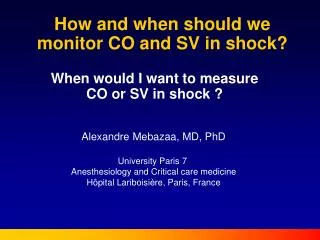
How and when should we monitor CO and SV in shock?. Alexandre Mebazaa, MD, PhD University Paris 7 Anesthesiology and Critical care medicine Hôpital Lariboisière, Paris, France. When would I want to measure CO or SV in shock ?. SHOCK MAP < 65mmHg Oliguria (<0.5ml/Kg/hour)

E N D
How and when should we monitor CO and SV in shock? Alexandre Mebazaa, MD, PhD University Paris 7 Anesthesiology and Critical care medicine Hôpital Lariboisière, Paris, France When would I want to measure CO or SV in shock ?
SHOCK MAP < 65mmHg Oliguria (<0.5ml/Kg/hour) Clinical signs of tissue hypoperfusion Volemia Heart function Vessel tone If shock is prolonged, mechanisms of shock are combined
Predominant RVF or global F PAC catheter Predominant LVF any CO monitoring SHOCK MAP < 65mmHg Oliguria (<0.5ml/Kg/hour) Clinical signs of tissue hypoperfusion • 1) Clinical approach • HR/BP • Peripheral perfusion • Impact of volume loading • Urine output First step Second step 2) CVP/SvcO2 3) Echocardiography should preceed any CO monitoring Third step Fourth step
Hemodynamic management of shock: first step- clinical evaluation
SHOCK MAP < 65mmHg Oliguria (<0.5ml/Kg/hour) Clinical signs of tissue hypoperfusion Heart rate Normal / high Heart rate < 40 bpm Give fluid challenge of 250 ml over 5 min Isoprenaline or pacemaker as necessary Yes, repeat if needed Improvement? No CVP/SvcO2
SvO2 >70% SvO2 <70% CVP low CVP high CVP N or low Hypovolaemic/ Haemorrhagic/ cause? Sepsis? Consider global/right ventricular failure Repeat fluid challenge (250ml/5mins) or transfusion if necessary. Repeat Fluid challenge 250ml/ 5mins Echocardiography that preceeds cardiac output monitoring Continue until normal values obtained Continue until normal values obtained Haemodynamic improvement ? No response Haemodynamic improvement Yes No Echocardiography that preceeds CO monitoring Vasopressors Insert CVP/SvcO2
Hemodynamic management of shock: third step- echocardiography
The « pyramid » of echocardiography skills in ICU Cholley,Vieillard-baron, Mebazaa, ICM 2006
Predominent right ventricular failure Predominent left ventricular failure Global heart failure TAMPONADE ? Massive mitral regurgitation ? Yes No No Echocardiographic guided pericardiocentesis or surgical intervention PA catheter LV dysfunction RV ischaemia? Pulmonary hypertension? Any CO Monitoring, ideally non invasive Reduce RV afterload, avoid excess volume, use inotropes if CO low Pulmonary vasodilators Optimise LV pre- and afterload, Inotropes if required Echocardiography Mebazaa et al. Intensive Care Med, 2004;30:185-96
Why/when would I want to measure CO or SV in shock? • Failure hemodynamic management based on clinical signs and CVP-ScvO2; this should always direct to echocardiography • Echocardiography should, ideally, always preceed CO monitoring • CO monitoring shoud be a PAC catheter in case of RV dysfunction while any CO monitoring, less invasive than PAC, should be favored for LV dysfunction
SHOCK MAP < 65mmHg Oliguria (<0.5ml/Kg/hour) Clinical signs of tissue hypoperfusion • 1) Clinical approach • HR/BP • Peripheral perfusion • Impact of volume loading • Urine output 2) CVP/SvcO2 3) Echocardiography should preceed any CO monitoring Predominant RVF or global F PAC catheter Predominant LVF any CO monitoring