Download
1 / 1
10 likes | 116 Vues
INTRODUCTION.

E N D
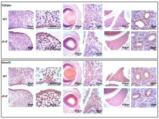
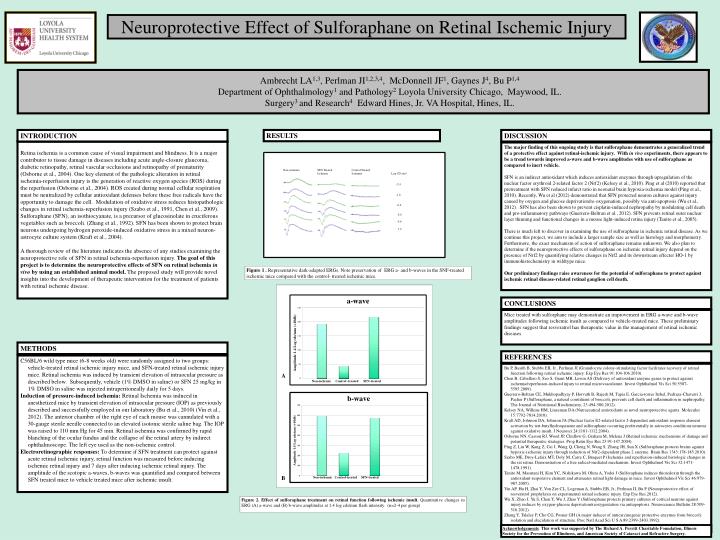
INTRODUCTION Retina ischemia is a common cause of visual impairment and blindness. It is a major contributor to tissue damage in diseases including acute angle-closure glaucoma, diabetic retinopathy, retinal vascular occlusions and retinopathy of prematurity (Osborne et al., 2004). One key element of the pathologic alteration in retinal ischemia-reperfusion injury is the generation of reactive oxygen species (ROS) during the reperfusion (Osborne et al., 2004). ROS created during normal cellular respiration must be neutralized by cellular antioxidant defenses before these free radicals have the opportunity to damage the cell. Modulation of oxidative stress reduces histopathologic changes in retinal ischemia-reperfusion injury (Szabo et al., 1991, Chen et al., 2009). Sulforaphane (SFN), an isothiocyanate, is a precursor of glucosinolate in cruciferous vegetables such as broccoli. (Zhang et al., 1992). SFN has been shown to protect brain neurons undergoing hydrogen peroxide-induced oxidative stress in a mixed neuron-astrocyte culture system (Kraft et al., 2004). A thorough review of the literature indicates the absence of any studies examining the neuroprotective role of SFN in retinal ischemia-reperfusion injury. The goal of this project is to determine the neuroprotective effects of SFN on retinal ischemia in vivo by using an established animal model. The proposed study will provide novel insights into the development of therapeutic intervention for the treatment of patients with retinal ischemic disease. DISCUSSION CONCLUSIONS The major finding of this ongoing study is that sulforaphane demonstrates a generalized trend of a protective effect against retinal-ischemic injury. With in vivo experiments, there appears to be a trend towards improved a-wave and b-wave amplitudes with use of sulforaphane as compared to inert vehicle. SFN is an indirect antioxidant which induces antioxidant enzymes through upregulation of the nuclear factor erythroid 2-related factor 2 (Nrf2) (Kelsey et al., 2010). Ping et al (2010) reported that pretreatment with SFN reduced infarct ratio in neonatal brain hypoxia-ischemia model (Ping et al., 2010). Recently, Wu et al (2012) demonstrated that SFN protected neuron cultures against injury caused by oxygen and glucose deprivation/re-oxygenation, possibly via anti-apoptosis (Wu et al., 2012). SFN has also been shown to prevent cisplatin-induced nephropathy by modulating cell death and pro-inflammatory pathways (Guerrero-Beltran et al., 2012). SFN prevents retinal outer nuclear layer thinning and functional changes in a mouse light-induced retina injury (Tanito et al., 2005). There is much left to discover in examining the use of sulforaphane in ischemic retinal disease. As we continue this project, we aim to include a larger sample size as well as histology and morphometry. Furthermore, the exact mechanism of action of sulforaphane remains unknown. We also plan to determine if the neuroprotective effects of sulforaphane on ischemic retinal injury depend on the presence of Nrf2 by quantifying relative changes in Nrf2 and its downstream effecter HO-1 by immunohistochemistry in wildtype mice. Our preliminary findings raise awareness for the potential of sulforaphane to protect against ischemic retinal disease-related retinal ganglion cell death. Mice treated with sulforphane may demonstrate an improvement in ERG a-wave and b-wave amplitudes following ischemic insult as compared to vehicle-treated mice. These preliminary findings suggest that resveratrol has therapeutic value in the management of retinal ischemic diseases. REFERENCES METHODS Bu P, Basith B, Stubbs EB, Jr., Perlman JI (Granulocyte colony-stimulating factor facilitates recovery of retinal function following retinal ischemic injury. Exp Eye Res 91:104-106.2010). Chen B, Caballero S, Seo S, Grant MB, Lewin AS (Delivery of antioxidant enzyme genes to protect against ischemia/reperfusion-induced injury to retinal microvasculature. Invest Ophthalmol Vis Sci 50:5587-5595.2009). Guerrero-Beltran CE, Mukhopadhyay P, Horvath B, Rajesh M, Tapia E, Garcia-torres Itzhel, Pedraza-Chaverri J, Pacher P (Sulforaphane, a natural constituent of broccoli, prevents cell death and inflammation in nephropathy. The Journal of Nutritional Biochemistry, 23:494-500.2012). Kelsey NA, Wilkins HM, Linseman DA (Nutraceutical antioxidants as novel neuroprotective agents. Molecules 15:7792-7814.2010). Kraft AD, Johnson DA, Johnson JA (Nuclear factor E2-related factor 2-dependent antioxidant response element activation by tert-butylhydroquinone and sulforaphane occurring preferentially in astrocytes conditions neurons against oxidative insult. J Neurosci 24:1101-1112.2004). Osborne NN, Casson RJ, Wood JP, Chidlow G, Graham M, Melena J (Retinal ischemia: mechanisms of damage and potential therapeutic strategies. Prog Retin Eye Res 23:91-147.2004). Ping Z, Liu W, Kang Z, Cai J, Wang Q, Cheng N, Wang S, Zhang JH, Sun X (Sulforaphane protects brains against hypoxic-ischemic injury through induction of Nrf2-dependent phase 2 enzyme. Brain Res 1343:178-185.2010). Szabo ME, Droy-Lefaix MT, Doly M, Carre C, Braquet P (Ischemia and reperfusion-induced histologic changes in the rat retina. Demonstration of a free radical-mediated mechanism. Invest Ophthalmol Vis Sci 32:1471-1478.1991). Tanito M, Masutani H, Kim YC, Nishikawa M, Ohira A, Yodoi J (Sulforaphane induces thioredoxin through the antioxidant-responsive element and attenuates retinal light damage in mice. Invest Ophthalmol Vis Sci 46:979-987.2005). Vin AP, Hu H, Zhai Y, Von Zee CL, Logeman A, Stubbs EB, Jr., Perlman JI, Bu P (Neuroprotective effect of resveratrol prophylaxis on experimental retinal ischemic injury. Exp Eye Res.2012). Wu X, Zhao J, Yu S, Chen Y, Wu J, Zhao Y (Sulforaphane protects primary cultures of cortical neurons against injury induces by oxygen-glucose deprivation/reoxygentation via antiapoptosis. Neuroscience Bulletin 28:509-516.2012) Zhang Y, Talalay P, Cho CG, Posner GH (A major inducer of anticarcinogenic protective enzymes from broccoli: isolation and elucidation of structure. Proc Natl Acad Sci U S A 89:2399-2403.1992). C56BL/6 wild type mice (6-8 weeks old) were randomly assigned to two groups: vehicle-treated retinal ischemic injury mice, and SFN-treated retinal ischemic injury mice. Retinal ischemia was induced by transient elevation of intraocular pressure as described below. Subsequently, vehicle (1% DMSO in saline) or SFN 25 mg/kg in 1% DMSO in saline was injected intraperitoneally daily for 5 days. Induction of pressure-induced ischemia: Retinal Ischemia was induced in anesthetized mice by transient elevation of intraocular pressure (IOP) as previously described and successfully employed in our laboratory (Bu et al., 2010) (Vin et al., 2012). The anterior chamber of the right eye of each mouse was cannulated with a 30-gauge sterile needle connected to an elevated isotonic sterile saline bag. The IOP was raised to 110 mm Hg for 45 min. Retinal ischemia was confirmed by rapid blanching of the ocular fundus and the collapse of the retinal artery by indirect ophthalmoscope. The left eye used as the non-ischemic control. Electroretinographic responses: To determine if SFN treatment can protect against acute retinal ischemic injury, retinal function was measured before inducing ischemic retinal injury and 7 days after inducing ischemic retinal injury. The amplitude of the scotopic a-waves, b-waves was quantified and compared between SFN treated mice to vehicle treated mice after ischemic insult. Neuroprotective Effect of Sulforaphane on Retinal Ischemic Injury Ambrecht LA1,3, Perlman JI1,2,3,4, McDonnell JF1, Gaynes J4, Bu P1,4 Department of Ophthalmology1 and Pathology2 Loyola University Chicago, Maywood, IL. Surgery3 and Research4 Edward Hines, Jr. VA Hospital, Hines, IL. RESULTS A Figure 1 . Representative dark-adapted ERGs. Note preservation of ERG a- and b-waves in the SNF-treated ischemic mice compared with the control- treated ischemic mice. B Non-ischemia SFN Treated Ischemia Control Treated Ischemia Log CD s/m2 -2.4 -1.6 -0.6 0.0 0.6 1.4 Figure 2.Effect of sulforaphane treatment on retinal function following ischemic insult. Quantitative changes in ERG (A) a-wave and (B) b-wave amplitudes at 1.4 log cds/mm flash intensity. (n=2-4 per group) Acknowledgements: This work was supported by The Richard A. Perritt Charitable Foundation, Illinois Society for the Prevention of Blindness, and American Society of Cataract and Refractive Surgery.