Download
1 / 140
1.45k likes | 2.08k Vues
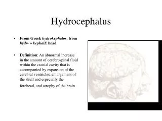
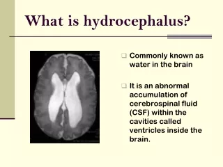
Hydrocephalus and Neuro Shunting. Sales Training April 2001. Hydrocephalus : From the Greek word hydro (water) & cephalo (head). A pathological condition where there is a disturbance in production, circulation and/or absorption of CSF, with subsequent accumulation of CSF

E N D
Hydrocephalus and Neuro Shunting Sales Training April 2001
Hydrocephalus:From the Greek • word hydro (water) & cephalo (head). • A pathological condition where there • is a disturbance in production, • circulation and/or absorption of CSF, • with subsequent accumulation of CSF • in the fluid-filled compartments of the • brain (ventricles).
About CSF(Cerebrospinal Fluid) • Clear, colorless fluid • Bathes, nourishes & protects brain and spinal cord. • Average CSF production-20ml/hr adults and 8ml/hr children • 400 to 500cc produced daily contains 15 to 45mg/100ml protein,some glucose, salts, urea and WBC’s
Ventricular System • Fluid filled cavities deep in cerebrum w/ pressure of 120-180mmH2O • Four ventricles • 2 Lateral • Third • Fourth • Connected by • Foramen of Monro • Aqueduct of Sylvius
Choroid Plexus Very vascular Found throughout but mostly in lateral Responsible for ICP waveform/ follows arterial pulse
Brain Layers/CSF Absorption A. - Arachnoid A.G. - Arachnoid Granulation B. - Bone C.A. - Cerebral Artery C.V. - Cerebral Vein D. - Dura Mater F.C. - Falx Cerebri P.M. - Pia Mater S. - Skin S.A.S. - Sub-Arachnoid Space S.D.S. - Sub-Dural Space S.S.S. - Superior Sagittal Sinus
CSF Flow-path • CSF flows in a caudal direction through the lateral, third and fourth ventricles • Exits through foramina of Luschka and Magendie into subarachnoid space around spinal cord and brain. • Absorption occurs through the arachnoid granulations into the venous system.
Communicating Non-communicating or Obstructive Normal Pressure Hydrocephalus Congenital Acquired Types of Hydrocephalus
CT Scan Showing severe hydrocephalus Normal CT Scan
Etiology of Hydrocephalus • Communicating • Overproduction/underabsorption of CSF • Choroid Plexus Papilloma-overproduces CSF • SAH • Infection • Neoplasms affecting the meninges • Trauma
Etiology of Hydrocephalus • Non-Communicating (Obstructive) • Aqueductal Stenosis • Arnold-Chiari Malformation (Cerebellar tonsils protrude into Foramen Magnum) • Cysts • Myelomeningocele • IVH • Tumors (particularly posterior fossa)
Normal Pressure Hydrocephalus • Usually present in elderly • Ventricular dilation despite normal CSF pressure • Triad of symptoms • 1) dementia • 2) gait disturbances (usually earliest) • 3) urinary incontinence
Signs & Symptoms Associated with Hydrocephalus • Infants • Increased head size • Bulging Fontanels • Separation of Cranial Sutures • Prominent Scalp Veins • Persistent Vomiting • Lethargy or irritability • “Setting Sun” eyes • Seizures • Delayed Development
S/S Associated with Hydrocephalus, cont. • Toddlers • Increased head size • Persistent vomiting • Headache • Lethargy or irritability • “Setting Sun” eyes • Blurred Vision • Seizures • Delayed Development
Hydrocephalus • “SETTING SUN” EYES
S/S Associated with Hydrocephalus, cont. • Older Children & Adults • Persistent Vomiting • Headache** • Visual Problems • Lethargy • Behavior Changes • Difficulty with schoolwork • Seizures
Diagnosis • Clinical Evaluation • Ultrasound (Intrauterine & through Fontanels. • CT Scan • MRI
Treatment Modalities • Surgical Procedures • Remove obstruction (Blood Clots, Tumors) • Endoscopic Third Ventriculostomy • Septal Fenestrations (Endoscopic) • Cyst Fenestrations (Endoscopic) • Shunt Insertion
Interventions for Hydrocephalus • If untreated: • *50-60% die of complications • If treated: • *40% normal intelligence • *70% live beyond infancy
Historical Treatment of Hydrocephalous • Hippocrates recognizes water accumulation in the brain. • 1545-Thomas Phaire-1st non-surgical treatment--Herbal plasters, head wraps • 18th Century--ventricular puncture--death from meningitis common • 1800’s-Variety of materials used to “wick” CSF from ventricles to subarachnoid space (i.e., linen threads, glass wool, rubber tube) • 1898-first lumboperitoneal shunt
Historical Treatment of Hydrocephalous, con’t • 1922-Dandy-third ventriculostomy through subfrontal • approach • 1923-Mixter-1st endoscopic 3rd Vent., choroid plexectomy • (L’Espinasse, Hildebrande, Dandy, Putnam and Scarff) • 1950’s-First effective CSF diversion with a one-way valve • using biocompatible synthetic materials. • John Holter-1st Silicone Valve • Robert Pudenz-Silicone distal slit valve • Peritoneum chosen as better absorptive site than the vascular system
Heyer Schulte and Shunt Industry History • 1953: Dr. Robert Pudenz and W.T. (Ted) Heyer team up on hydrocephalus research • 1955: Pudenz ventriculo-atrial shunt is developed • 1959: Rudy Schulte joins Heyer and Pudenz • 1959: Pudenz flushing valve is developed • 1960: Codman distributes Heyer-Schulte products • 1960: Holter valve is created • 1965: Cordis begins U.S. presence • 1965: Extra-Corporeal buys Holter • 1973: Codman dropped as Heyer-Schulte distributor
Heyer Schulte and Shunt Industry History • 1974: American Hospital Supply buys Heyer-Schulte • 1975: Codman introduces their own product line • 1977: Anasco, PR manufacturing facility is built • 1978: Codman buys Extra-Corporeal • 1983: AHS folds Heyer-Schulte into V. Mueller • 1984: Dr. Pudenz and Rudy Schulte found P-S Medical • 1986: Baxter-Travenol acquires AHS
Heyer Schulte and Shunt Industry History • The 90’s • NeuroCare Group acquires Heyer-Schulte • Radionics introduces full shunt line • Medtronic acquires P-S Medical • Phoenix Biomedical enters the market • Codman acquires Cordis • Elekta acquires Cordis • NMT acquires Cordis • Integra acquires Heyer-Schulte
What is a Shunt? • A shunt is a device that diverts CSF from the CNS (usually the lateral ventricle or the lumbar subarachnoid space) to an alternate body cavity (usually the peritoneum or the right atrium) where it is reabsorbed.
How Shunts Work • Divert CSF from the CNS to another body cavity (R atrium, peritoneum) for absorption. • Mechanical device that regulates flow out of the ventricle. • One-way valve opens when the sum of the forces acting on it exceed some threshold. (the difference between the inlet or ventricular pressure and outlet or peritoneal pressure.
Shunt Systems • Ventriculo-peritoneal • Ventriculo-atrial • Lumbar-peritoneal
Shunt Components • Primary Components • Proximal Catheter • Valve (Proximal or Distal) • Distal Catheter • Optional Components • Reservoir • Siphon Limiting Mechanism (ASD, SCD, GCD) • Accessories • Connectors • Guides • Introducers/Stylets • Catheter Passers
SHUNT ACCESSORIES • Proximal catheter stylet (can use endoscope) • Stylets for unitized shunts • Shunt passers • Connectors and Right angle guides • Shunt tap kits • Manometers
Valve Mechanisms • Differential Pressure Valves • Flow regulating devices
Valve Mechanisms • Differential Pressure Valves • Valves open when difference between the ventricular pressure and the peritoneal pressure exceeds some threshold. • Pressure difference at which a valve opens is called the opening pressure. • Pressure difference at which a valve closes is called the closing pressure.
Valve Types • Burr Hole - shaped to fit the hole made in the skull. • The reservoir is an integral part e.g. Pudenz • Flat Bottom - rests flat against the skull distal to the • ventricular catheter e.g. LPV II, Novus • Cylindrical/In Line - appears “seamless” between the • ventricular and peritoneal catheters • e.g.. Ultra VS