Download
1 / 25
260 likes | 411 Vues
HEART BLOCKS. Josali Sandy G. Dy. Sinoatrial Block. Sinoatrial Exit Block Periodic absence of a P wave Type I ( Weckenbach ): Progressive shortening of the P-P interval followed by a long P-P cycle that is shorter than the sum of any two consecutive P-P intervals.

E N D
HEART BLOCKS Josali Sandy G. Dy
Sinoatrial Block • Sinoatrial Exit Block • Periodic absence of a P wave • Type I (Weckenbach): Progressive shortening of the P-P interval followed by a long P-P cycle that is shorter than the sum of any two consecutive P-P intervals. • Type II: The long P-P intervals are exact whole-number multiples of the shortest constant P-P interval.
PP intervals gradually shorten until a pause occurs (i.e., the blocked sinus impulse fails to reach the atria) • The pause duration is less than the two preceding PP intervals • The PP interval following the pause is greater than the PP interval just before the pause
PP intervals fairly constant (unless sinus arrhythmia present) until conduction failure occurs. • The pause is approximately twice the basic PP interval
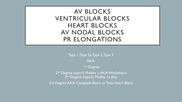
AV Nodal Blocks • First Degree AV Block • P-R interval greater than 0.21 second • One to one atrioventricular conduction.
AV Nodal Blocks • Second Degree AV Blocks • Presence of sinus rhythm. • Some P waves are followed by QRS complexes. Others are not. • Mobitz type I: P-R intervals of conducted beats vary according to Weckenbach periodicity; increasing P-R intervals. • Mobitz type II (Hay): P-R intervals of conducted beats are normal or prolonged but constant.
Th AV Nodal Blocks • Third Degree AV Block/ Complete Heart Block • Atrioventricular dissociation (p waves are seen marching through the QRS complexes) • P-P interval is less than the R-R interval • Idioventricular rhythm
Intraventricular Block • Right Bundle Branch Block • QRS duration is 0.12 second or longer • The QRS complex in lead V1 has an rsR’ configuration or is a solitary R wave.
Intraventricular Block • Left Bundle Branch Block • QRS duration is 0.12 second or longer • The QRS complex is notched and splintered and has QS or rS deflection in lead V1
LBBB RBBB
Intraventricular Block • Non-specific Intraventricular Block • QRS complex duration exceeding 0.10 second. • Absence of typical ECG characteristics of right bundle branch block and left bundle branch block. • Causes of nonspecific IVCD's include: • Ventricular hypertrophy (especially LVH) • Myocardial infarction (so called periinfarction blocks) • Drugs, especially class IA and IC antiarrhythmics (e.g., quinidine, flecainide) • Hyperkalemia
Fascicular Block • Left Anterior Fascicular Block (LAFB) - the most common intraventricular conduction defect • Left axis deviation in frontal plane, usually -45 to -90 degrees • rS complexes in leads II, III, aVF • Small q-wave in leads I and/oraVL • R-peak time in lead aVL >0.04s, often with slurred R wave downstroke • QRS duration usually <0.12s unless coexisting RBBB Usually see poor R progression in leads V1-V3 and deeper S waves in leads V5 and V6 • May mimic LVH voltage in lead aVL, and mask LVH voltage in leads V5 and V6.
Fascicular Block • Left Posterior Fascicular Block (LPFB) - Very rare intraventricular defect • Right axis deviation in the frontal plane (usually > +100 degrees) • rS complex in lead I • qR complexes in leads II, III, aVF, with R in lead III > R in lead II • QRS duration usually <0.12s unless coexisting RBBB • Must first exclude (on clinical grounds) other causes of right axis deviation such as corpulmonale, pulmonary heart disease, pulmonary hypertension, etc., because these conditions can result in the identical ECG picture!
Fascicular Block • Bifascicular Blocks • RBBB plus either LAFB (common) orLPFB (uncommon) • Features of RBBB plus frontal plane features of the fascicular block (axis deviation, etc.)
ECG shows classic RBBB (note rSR' in V1) plus LAFB (note QRS axis = -45 degrees, rS in II, III, aVF; and small q in aVL)
Fascicular Block • Trifascicular Block • Any of the following combinations may be present: • RBBB + LAHB + AV Block • RBBB+ LPHB+AV Block • prolongation of the PR interval (first degree AV block) • right bundle branch block • either left anterior fascicular block or left posterior fascicular block
SINUS ARREST • A sinus rhythm is interrupted by a sudden lengthening of the P-P cycle. • The long P-P interval varies in duration and is not a whole number multiple of the basic sinus cycle and the shortest of such intervals is usually slightly less than twice the basic sinus cycle.