Download
1 / 68
760 likes | 2.01k Vues
Review of Cranial and Spinal Nerves. Randy Perkins, PhD Department of Physical Therapy and Human Movement Sciences Northwestern University Feinberg School of Medicine.

E N D
Review of Cranial and Spinal Nerves Randy Perkins, PhD Department of Physical Therapy and Human Movement Sciences Northwestern University Feinberg School of Medicine
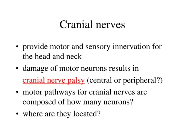
1. A patient who has been exhibiting various endocrine abnormalities has an MRI scan of the head. This scan reveals a small tumor of the pituitary gland. As this tumor expands superiorly what visual field defect will this patient exhibit? A. Left or right monocular blindness B. Binasal hemianopsia C. Left or right homonymous hemianopsia D. Bitemporal hemianopsia
VISUAL FIELDS Temporal Nasal Temporal Answer: D. Bitemporal hemianopsia is caused by compression of the nasal fibers crossing in the optic chiasm. Nasal Temporal
2. A 49 year old woman is in a motor vehicle accident and sustains a closed head injury. A CT scan does not show any intracranial hemorrhage but reveals a small tumor at the cerebellopontine angle of the brain. Which of the following nerves is most likely to be affected by this tumor? A. Facial nerve B. Glossopharyngeal nerve C. Abducens nerve D. Trigeminal nerve E. Vagus nerve
Answer: A. Facial nerve will be compressed by an acoustic neuroma of the vestibulocochlear nerve.
3. A man is involved in an MVA and sustains trauma to the middle ear, damaging the chorda tympani. Salivation from which of the following will be affected? A. Parotid, submandibular and sublingual glands B. Submandibular gland only C. Sublingual gland only D. Parotid gland only E. Submandibular and sublingual glands
Answer: E. Submandibular and sublingual glands are innervated by the parasympathetic fibers traveling in the chorda tympani branch of the facial nerve.
4. A physician is performing a cranial nerve exam on a patient. While testing the gag reflex it is noted that when the left side of the pharyngeal mucosa is touched, the patient gags and his uvula deviates to the left. When the right side is touched, the patient does not gag. Which of the following is the most likely location of his lesion? A. Left glossopharyngeal and vagus nerves B. Right glossopharyngeal and vagus nerves C. Right glossopharyngeal nerve only D. Right vagus nerve only E. Left glossopharyngeal nerve only
Answer: B. Right glossopharyngeal (sensory limb of gag reflex) and vagus (motor limb of gag reflex) nerves. The uvula deviates to the left because the right levator veli palatini muscle is paralyzed.
Clinical test of the vagus nerve: “Say Ah.” In a lower motor neuron lesion of the vagus nerve the uvula deviates away from the lesion side (away from the weak side). Nucleus ambiguus LMN lesion Vagus nerve (Fix, 2nd ed., p. 73)
5. During a physical exam, the physician asks the patient to say “Kuh, kuh, kuh,” “La-la-la,” and “Mi-mi-mi.” The patient is unable to say “la” but says the other two sounds correctly. A lesion of which of the following nerves is most strongly suggested by these findings? A. VII B. IX C. XII D. X E. V
Answer: C. XII. Saying “la” requires the tongue to be placed against the roof of the mouth.
6. A 54 y. o. woman presents with complaints of double vision. While testing the patient’s eye movements, the physician notes that the patient cannot elevate her right eye from the abducted position. Which of the following muscles is paralyzed? A. Inferior oblique B. Inferior rectus C. Medial rectus D. Superior rectus E. Superior oblique
Answer: D. Superior rectus can elevate and adduct the eye from the neutral position. From the abducted position it is the only muscle that can elevate the eye.
7. As part of a complete neurological exam a medical student touches the patient’s right eye with a thin wisp of cotton as the patient looks to the left. The patient closes both of his eyelids in response. Which of the following cranial nerves is responsible for the motor limb of this reflex? A. Facial B. Abducens C. Oculomotor D. Trigeminal E. Trochlear
Answer: A. Facial nerve is the motor limb of the corneal (blink) reflex, causing bilateral contraction of the orbicularis oculi muscles.
The facial motor nucleus receives fibers from the spinal trigeminal sensory nucleus for the bilateral corneal (blink) reflex. Irritation of one cornea will cause both eyes to blink. (Fix, 2nd ed., p. 68)
8. A 46 y. o. woman presents to her physician with complaints of double vision. She is unable to adduct her right eye on attempted left lateral gaze. Convergence is intact. Both the direct and consensual light reflexes are normal. Which of the following structures is most likely affected? A. Left oculomotor nerve B. Medial longitudinal fasciculus C. Right abducens nerve D. Right oculomotor nerve E. Left abducens nerve
Answer: B. Medial longitudinal fasciculus. The right MLF has been damaged, most likely by a demyelinating plaque seen in multiple sclerosis. This is termed internuclear ophthalmoplegia or INO.
Lateral gaze is controlled by the paramedian pontine reticular formation (PPRF) in the lateral (parvocellular) reticular area located near the abducens nucleus. The PPRF sends fibers to the ipsilateralabducens nucleus and, through the medial longitudinal fasciculus (MLF), to the contralateraloculomotor nucleus, thereby coordinating the actions of the lateral and medial recti during lateral gaze. III MLF PPRF VI (Kandel et al., 4th ed., p. 791)
9. An otherwise healthy 11 y. o. female with recurrent upper respiratory tract infections undergoes bilateral tonsillectomy. While performing the procedure, the surgeon accidentally damages the nerve that lies in the tonsillar fossa deep to the palatine tonsil. Which of the following functional losses is likely to result from this injury? A. Loss of sensation on the posterior ⅓ of the tongue B. Loss of taste on the anterior ⅔ of the tongue C. Paralysis of the constrictor muscles of the pharynx D. Paralysis of the muscles of the soft palate E. Paralysis of the tongue muscles
Answer: A. Loss of sensation on the posterior ⅓ of the tongue because the glossopharyngeal nerve was damaged.
10. To evaluate hypoglossal nerve function, a physician asks her patient to protrude his tongue. On doing so, his tongue deviates to the right side. This finding results from paralysis of which of the following muscles? A. Left genioglossus B. Left hyoglossus C. Right genioglossus D. Right hyoglossus E. Left styloglossus
Answer: C. Right genioglossus paralysis will cause the tongue to deviate toward the right when protruded. The tongue always deviates toward the weak/paralyzed side when protruded.
Clinical test of the hypoglossal nerve: “Stick out your tongue.” In a lower motor neuron lesion of the hypoglossal nerve the tongue deviates to the lesion side (toward the weak side) when protruded. Hypoglossal nucleus LMN lesion Hypoglossal nerve (Fix, 2nd ed., p. 76)
11. A patient arrives in the ER after having suffered severe head trauma in a motorcycle accident. Radiographic studies of the head reveal a basilar skull fracture in the region of the foramen ovale. Which of the following functional losses would most likely be related to this injury? A. Loss of abduction of the eye B. Loss of sensation over the forehead C. Loss of taste on the anterior ⅔ of the tongue D. Loss of sensation over the zygoma E. Paralysis of muscles of mastication
Answer: E. Paralysis of muscles of mastication caused by damage to the mandibular division of the trigeminal nerve as it exits foramen ovale.
12. A patient presents with complaints of chronic headaches. A cerebral arteriogram reveals a large aneurysm of the right superior cerebellar artery immediately distal to its origin from the basilar artery. Which of the following findings is most likely to be seen as a result of compression of a cranial nerve by this aneurysm? A. Loss of abduction of the right eye B. Loss of adduction of the right eye C. Loss of depression of the right eye from the adducted position D. Loss of sensation from the right side of the face E. Loss of vision in the right eye
Answer: B. Loss of adduction of the right eye caused by compression of the oculomotor nerve Oculomotor nerve Superior cerebellar artery
13. A 24 y. o. woman presents to her physician with an inability to close her right eye. Physical exam reveals weakness of the right orbicularis oculi. Which of the following symptoms would likely also be present? A. Double vision B. Inability to feel the face C. Inability to chew D. Hyperacusis E. Inability to shrug the shoulder
Answer: D. Hyperacusis (sensitivity to loud sounds) caused by paralysis of the stapedius muscle innervated by the facial nerve.
14. A patient presents with pain radiating down the S1 dermatome on the left side, weakness of muscles in the S1 myotome on the left side and loss of the ankle jerk reflex on the left side. What is the most likely cause of these symptoms? A. Complete spinal cord transection at S1 B. Left hemisection of the cord at S1 C. Compression of the S1 left dorsal root D. Compression of the S1 left ventral root E. Compression of the S1 left dorsal and ventral roots
Answer: E. Compression of the S1 left dorsal and ventral roots caused by herniation of the L5-S1 disc.
15. A lesion of the brachial plexus affecting the two nerves indicated by the arrows at the upper right in the diagram would result in: A. Loss of sensation along the medial side of the forearm and hand B. Loss of flexion, abduction and lateral rotation of the arm C. Hyperextension of the MCP joints and flexion of the IP joints D. Loss of thumb opposition E. Loss of wrist extension
Answer: B. Loss of flexion, abduction and lateral rotation of the arm (C5 and C6 upper plexus injury, waiter’s tip hand).
16. A lesion of which branch of the brachial plexus will cause difficulty in using crutches? A. B. C. D. E.
Answer: D. Thoracodorsal nerve (nerve to latissimus dorsi) extends, adducts and medially rotates the arm at the glenohumeral joint.
17. Following a brachial plexus injury a patient presents with a claw-hand deformity. In what areas of skin would you expect to see anesthesia or loss of sensation? A. Medial side of the hand and medial 1 ½ digits B. C5, 6 dermatomes C. Lateral side of the hand and lateral 3 ½ digits D. C8, T1 dermatomes E. Lateral side of the arm and forearm
Answer: D. C8, T1 dermatomes (lower plexus injury).
Lower brachial plexus injury (Klumpke’s palsy) • involves stretching of the C8 and T1 roots or lower trunk of the brachial plexus and • is caused by a forceful upward pull of the shoulder. C8 T1 Augusta Dejerine Klumpke 1859-1927 (Moore & Agur, 2nd ed., p. 440)
Effects of injury: • the paralysis and anesthesia primarily affect the muscles and skin supplied by the ulnar nerve. • Wrist and finger movements are most affected, and a claw-hand results. C8 T1 Ulnar nerve (Moore & Agur, 2nd ed., p. 440)
There is anesthesia in the C8 and T1 dermatomes along the medial side of the forearm and hand. C8 T1 (Moore & Agur, 2nd ed., p. 420)
18. A Northwestern running back hurt his leg in a game and used axillary crutches for several weeks to protect it while it healed. He then complained that abduction of his arm had become weak and so had extension of his elbow. Flexion of his elbow and arm seemed to be normal, as did flexion of his wrist and movement of his fingers. You diagnose injury to his: A. Lower trunk of the brachial plexus B. Medial cord of the brachial plexus C. Posterior cord of the brachial plexus D. Lateral cord of the brachial plexus E. Axillary nerve
Answer: C. Posterior cord of the brachial plexus (axillary nerve produces abduction of the arm and the radial nerve produces extension of the elbow).
19. Following breast surgery a patient complained that she was having difficulty raising her arm above her head to comb her hair. Furthermore, the medial edge of her scapula was noticeably lifted away from the thoracic wall. Most likely, the surgeon had injured which of the following structures? A. Long thoracic nerve B. Thoracodorsal nerve C. Upper trunk of the brachial plexus D. Axillary nerve E. Lower trunk of the brachial plexus
Answer: A. Long thoracic nerve (nerve to serratus anterior) injury produces winging of the scapula. Serratus anterior protracts and rotates the scapula upward.
20. A patient presents with loss of abduction and weakness of lateral rotation of the arm and anesthesia over the lateral surface of the shoulder. These symptoms could result from a nerve injury caused by: A. Mastectomy operation B. Fracture of the humeral surgical neck C. Forceful upward pull of the shoulder during birth D. Fracture of the humeral shaft E. Pronator syndrome
Answer: B. Fracture of the humeral surgical neck can cut the axillary nerve that innervates deltoid and teres minor. Axillary nerve
21. A patient presents with loss of sensation on the radial half of the back of the hand. What deformity of the hand would you expect this patient to have? A. Ape hand B. Waiter’s tip hand C. Claw-hand D. Sign of benediction E. Wrist drop
Answer: E. Wrist drop (radial nerve).