Download
1 / 10
100 likes | 187 Vues
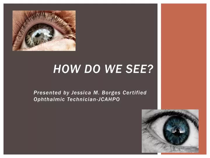
How do we see?. Presented by Jessica M. Borges Certified Ophthalmic Technician-JCAHPO. An Introduction into Optics.

E N D
How do we see? Presented by Jessica M. Borges Certified Ophthalmic Technician-JCAHPO
An Introduction into Optics • Many of us when we go to a Doctors office no matter what the specialty do not fully understand what is being performed on us, or why our body reacts in certain ways that bring us to see the doctor to begin with. • Our eyes, vital organs of our central nervous system are complex comprised of different structures all working together to give us our sight. • Eye Doctors examine our eyes for disease, and perform diagnostic tests to corrective for any refractive errors (difficulty with vision due to the shape of our eyes) we may possess. • My goal is to educate you on how our eyes work, how it is we see and why the tests an Ophthalmologist or Optometrist performs are of importance.
The shape of your Cornea Influences How Light Enters your Eye and how you see. • Hyperopia -Farsightedness; Distance is Clear and your Near Vision is Blurry. Occurs when your eyeball is too short, so light rays enter the eye and go beyond the “perfect point” and into your choroid. • Cornea – The First part of the eye’s optical system. This transparent membrane begins the process of focusing light the eye receives. Depending on its shape the light will reach its destination, scatter and get lost or go farther then needed for clear vision.
Refractive Errors • Astigmatism- Cause by an Irregularly shaped Cornea (similar to a football shape) or a curvature to the lens on your eye. Causes blurry vision with Distance and sometimes Near depending on the degree of astigmatism.. • Myopia-Nearsightedness; Your near vision is clear and your distance vision is blurry. Occurs when the eyeball is too long, so light rays enter the eye and scatter and do not make it to the “perfect point” for clear distance vision.
You have a lens similar to a contact lens or glasses lens that is suspended by muscles in your eye that further assist in the refraction of light • Attached to the back of the colored part of your eye (your Iris) attached by muscles is your natural crystalline lens which helps to further direct light rays into the back of your eye. • As you age the muscles become weaker and the lens becomes thicker and harder with age (Presbyopia) gets cloudy(developing into a cataract) and this aging alters how light rays enter (or get stopped) and how you can see.
Presbyopia and Cataracts • Clouding caused by Cataract
The Retina is a light sensitive Tissue in the back of your eye that assists images being transmitted to your brain. • The retina is the light sensitive tissue that receives light and turns that light into electrical impulses that trigger nerve impulses to send images through the optic nerve to your brain. • The dark pigmented spot is your macula which surrounds a small dimple called the fovea that is responsible for the majority of your color vision and your sharpest vision. Individuals who acquire macular degeneration end up with a loss of their central vision. • The optic nerve is the whitish disk and your eyes natural blind spot. The optic nerve sends images to the brain and is what gets affected in patients that have glaucoma (loss of peripheral vision due to increased pressure in the eye damaging the optic disk.)
Fundus Photo of Retina, Macula, Optic Nerve This photo is of a healthy retina (orange tissue), macula (dark spot) and optic nerve (yellow/whitish disk)
Conclusion • How we see is of vital importance to us, and our visual organs are very complex, but essentially all of our sight comes from three components. • The Cornea, the lens and the retina make up our optical system and they each assist light rays in their transmission to the brain who processes those rays as images. • Depending on the shape of our eye and whether we are corrected properly for that refractive error will determine how clearly we see objects around us. • Having an eye exam regularly is important to asses the health of our eye and to correct for any changes that may occur as we age.
Reference Page • M.D.,Stein,H.A., M.D.,Slatt, B.J., & M.D.,Stein, R.M. (2000)The Ophthalmic Assistant: A Guide for Ophthalmic Medical Personnel(7th ed.).San Francisco, CA. American Academy of Ophthalmology. • M.D.,Carr, Tyree. (1999) Ophthalmic Medical Assisting; An Independent Study Course(3rd ed.). San Francisco, CA. American Academy of Ophthalmology. • Ledford, Janice. (2004)Certified Ophthalmic technician Exam Review Manual (2nd ed.). Thorofare, NJ. SLACK Incorporated. • M.D., Wilson, Fred. (1996)Practical Ophthalmology A Manual for Beginning Residents.(4th ed.)San Francisco, CA. American Academy of Ophthalmology. • Elkington, A. R. ; Khaw, P. T. (1988)Refractive Errors. British Medical Journal, Vol.297(6642), pp.192-195