Download

1 / 6
60 likes | 167 Vues
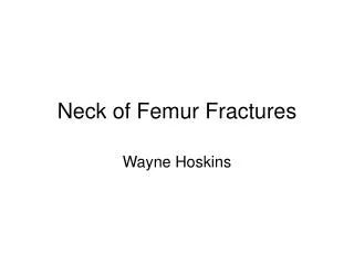
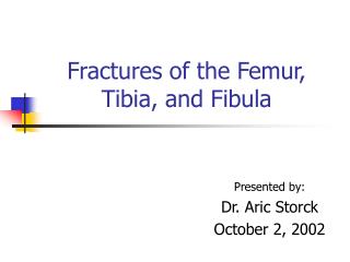
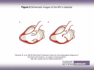
AC HUOL 260412 Figure 1. Images of the Left Shoulder and Femur.

E N D
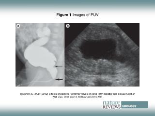
AC HUOL 260412Figure 1. Images of the Left Shoulder and Femur. A chest radiograph (Panel A) shows a sharply defined lucent lesion in the left scapula (arrow). A scan from combination positron-emission tomography and CT performed 6 weeks earlier shows a sharply defined lytic lesion with a “punched-out” appearance (Panel B, arrow) and intense 18F-fluorodeoxyglucose uptake in the lesion (Panel C). An axial unenhanced CT scan of the distal shaft of the right femur obtained at the time of the CT-guided fine-needle aspiration biopsy (Panel D) shows a sharply defined lucent defect in the cortex of the femur. The lesion has a punched-out appearance, with margins that resemble those of the left scapular lesion.
AC HUOL 260412 A B
AC HUOL 260412 C D
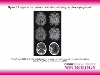
AC HUOL 260412 A B
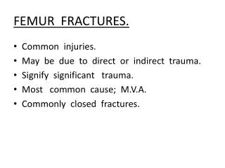
AC HUOL 260412Figure 2. Images of the Lungs C A chest radiograph obtained at the time of admission to this hospital (Panel A) shows subtle, fine reticulonodular opacities in both lungs that appear to spare the costophrenicsulci. An axial CT scan of the chest (Panel B) shows small, irregular pulmonary nodules that measure 2 to 3 mm in diameter; some nodules appear centrilobularin distribution. Several nodules appear to be cavitating, and there are small central areas of low attenuation. Small cysts of various sizes are also seen in both lungs. The small nodules and cysts predominantly involve the upper and middle zones of the lungs and appear to spare the lung bases and costophrenicsulci. This distribution of findings is more apparent on a reformatted coronal CT image (Panel C).