Download
1 / 12
130 likes | 382 Vues
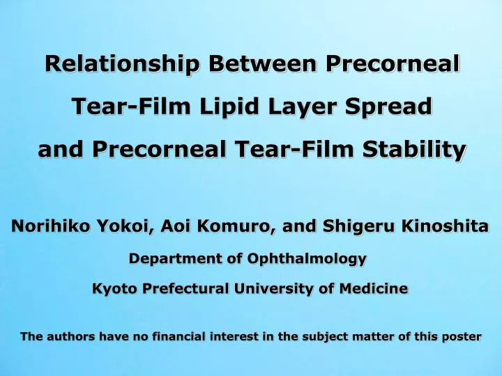
Relationship Between Precorneal Tear-Film Lipid Layer Spread and Precorneal Tear-Film Stability. Norihiko Yokoi, Aoi Komuro, and Shigeru Kinoshita Department of Ophthalmology Kyoto Prefectural University of Medicine.

E N D
Relationship Between Precorneal Tear-Film Lipid Layer Spread and Precorneal Tear-Film Stability Norihiko Yokoi, Aoi Komuro, and Shigeru Kinoshita Department of Ophthalmology Kyoto Prefectural University of Medicine The authors have no financial interest in the subject matter of this poster
Purpose Dynamics of the Tear-Film Lipid Layer (TFLL) spread after opening the eye follows a rheological Voigt model (Yokoi N, et al. IOVS2008;49), and the initial velocity of the TFLL spread [H´(0), mm/sec.] provides the quantitative value for the tear volume, both in theory and in practice. The elapsed amount of time from the opening of the eye until the breakup (Non-invasive breakup time: NIBUT, sec.) of the precorneal tear filmis an indication of tear film stability (Ishibashi T, Yokoi N, et al. J Glaucoma 2003;12). Therefore, we investigated the relationship between H´(0) and NIBUT.
Subjects (39 eyes from39 cases) Males:3 eyes from 3 cases; Females:36 eyes from 36 cases Normal eyes : 4 eyes from 4 cases Definite dry eye : 20 eyes from 20 cases Suspected dry eye: 12 eyes from 12 cases Post punctal occlusion : 3 eyes from 3 cases (diagnoses based on 2006 dry eye diagnostic criteria in Japan) No meibomian gland dysfunction Mean Age: 52.1±14.8 (SD) yrs.
Methods Investigation of the correlation between: ①, ②, ③ Video-Meniscometry Video-Interferometry ① ( ) ② ③ ( ) ① Measurement ofR(Radius of the central lower tear meniscus) ② Measurement ofH’(0)(Initial velocity of the TFLL spread) ③ Measurement ofNIBUT(Non-invasive breakup time) of the precorneal tear film Note: In cases of severe tear deficiency where the TFLL cannot be seen ⇒H’(0)=0mm/sec. and NIBUT=0 sec. In cases of stable tear film where no NIBU is seen at 10 seconds ⇒ NIBUT=10 sec.
Automatic analysis of the spreading TFLL using the Voigt model and the cross-correlation method • A certain rectangular area isdetermined on a captured image of the • spreading lipid layer [Image (A)]. • A rectangular area of the same size and having the closest total brightness to that in Image (A) is automatically searched for in Image (B) 0.05 seconds after Image (A) based on the cross-correlation method using a custom made computer program. 3) The transitional shift along the y-axis(dy) of the rectangular area between (A) and (B) is then calculated; dyis subsequently summed over time and adopted to the Voigt model. (B) (A) dy
Analysis of the TFLL spread along the y-axis using the Voigt-model equation Voigt-model equation: H = ρ [1-e (-t /λ) ] H: summed shift of the rectangular area along the y-direction ρ: constant, t: time, λ: retardation time=ρ(1-1/e) H’(0) (initial velocity of the TFLL spread along the y-axis) was used as a parameter for evaluating the TFLL spread
Representative measurement of H’(0) (Left figure) and NIBUT (Right figure)of the precorneal tear film Before opening of the eye Sum of the upward spread of the TFLL along the Y-axis (mm) Opening of the eye t=1 sec. t=0 sec. H´(0)=10.8 mm/sec. 4.0 t=1.6 sec. t=3 sec. t=4 sec. 3.0 Actual value 2.0 t=5 sec. t=5.8 sec. t=7 sec. Voigt model NIBU 1.0 t=8 sec. t=9 sec. t=10 sec. 0.0 0.2 0.4 0.6 0.8 1.0 1.2 1.4 1.6 1.8 2.0 Time (sec.) NIBUT=5.8 sec
Results (1) Relationship between R and H’(0) 12 10 rs=0.6751 8 (Spearman) 6 H’(0) (mm/sec.) P<0.0001 4 2 0 n=39 -2 0 .1 .2 .3 .4 .5 .6 .7 .8 1.0 R (mm)
Results (2) Relationship between H’(0) and NIBUT 12 10 rs=0.9039 (Spearman) 8 P<0.0001 NIBUT (sec.) 6 4 2 0 n=39 -2 -2 8 10 12 0 2 4 6 H’(0) (mm/sec.)
Results (3) Comparison of the NIBUT using the average (=3.3mm/sec.) of H’(0) as the cut-off value 12 *P<0.0001 * (Student’s t-test) 10 mean±SD 8 NIBUT (sec.) 6 4 (20) 2 (19) 0 H’(0)≧3.3 H’(0)<3.3
Summary of the Results Using a custom made computer program for cross-correlation in combination with the Voigt model, the initial velocity of the TFLL spread along the y-axis [H’(0)] was measured. A significant positive relationship was seen between the radius of tear meniscus (R) and H’(0) (rs=0.6751; P<0.0001). 1) There was a significant positive relationship between H’(0) and the non-invasive breakup time(NIBUT) of the precorneal tear film (rs=0.9039; P<0.0001). 2) Using the average (=3.3mm/sec.) of the initial velocity of the TFLL spread in 39 subject eyes as the cut-off value, NIBUT in the H’(0) < 3.3group was significantly shorterthan in the H’(0) ≧ 3.3 group (P<0.0001). 3)
Conclusion Based on the findings of Berger & Corrsin (J Biomech 1974;7) regarding the significant correlation between the velocity of the TFLL spread and the precorneal tear-film aqueous-layer thickness, as well as the findings of the present study showing a significant correlation between the velocity of the TFLL and the radius of the tear meniscus, it may be concluded that the precornealtear-film aqueous-layer thickness contributes to the stability of the precorneal tear film.