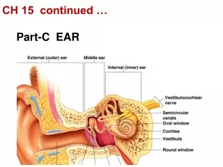
Download

1 / 35
350 likes | 459 Vues
Continued from part a. Also Raman. Not Raman, unless RR. Weak IR Multiple bands. Peptide conformation depends on f , y angles. If ( f,y) repeat, they determine secondary structure. Chromophores – amides are locally achiral CD has little signal without coupling, ideal for detection

E N D
Also Raman Not Raman, unless RR Weak IR Multiple bands
Peptide conformation depends on f, y angles If (f,y) repeat, they determine secondary structure Chromophores – amides are locally achiral CD has little signal without coupling, ideal for detection -- IR, Raman resolve shift Detection requires method sensitive to amide coupling Far UV absorbance broad, little fluorescence—coupling impact small
I II Model polypeptideIR absorbancespectra - Amide I and II (Not in Raman) (weak IR but strong in Raman)
Combining Techniques: Vibrational CD “CD” in the infrared region Probe chirality of vibrations goal stereochemistry Many transitions / Spectrally resolved / Local probes Technology in place -- separate talk Weak phenomenon - limits S/N / Difficult < 700 cm-1 Same transitions as IR same frequencies, same resolution Band Shape from spatial relationships neighboring amides in peptides/proteins Relatively short length dependence AAn oligomers VCD have DA/A ~ const with n vibrational (Force Field) coupling plus dipole coupling Development -- structure-spectra relationships Small molecules – theory / Biomolecules -- empirical, Recent—peptide VCD can be simulated theoretically
VIBRATIONAL OPTICAL ACTIVITY Differential Interaction of a Chiral Molecule with Left and Right Circularly Polarized Radiation During Vibrational Excitation VIBRATIONAL CIRCULAR DICHROISMRAMAN OPTICAL ACTIVITY Differential Absorption of Left and Right Differential Raman Scattering of Left Circularly Polarized Infrared Radiation and Right Incident and/or Scattered Radiation
UIC Dispersive VCD Schematic Yes it still exists and measures VCD! Electronics Optics and Sampling
Separate VCD Bench Optics FTIR UIC FT-VCDSchematic (designed for magnetic VCD commercial ones simpler) Electronics Polarizer PEM (ZnSe) Sample detector FT-computer Optional magnet Detector (MCT) filter lock-in amp PEM ref
ma Tab mb Large electric dipole transitions can couple over longer ranges to sense extended conformation Simplest representation is coupled oscillator De = eL-eR Dipole coupling results in a derivative shaped circular dichroism l Real systems - more complex interactions - but pattern is often consistent
Amide I Amide II DA a b coil Selected model Peptide VCD, aqueous solution
Nature of the peptide random coil form Tiffany and Krimm in 1968 noted similarity of Proline II and poly-lysine ECD and suggested “extended coil” Problem -- CD has local sensitivity to chiral site --IR not very discriminating Dukor and Keiderling 1991 with ECD, VCD, and IR showed Pron oligomers have characteristic random coil spectra Suggests -- local order, left-handed turn character -- no long range order in random coil form Same spectral shape found in denatured proteins, short oligopeptides, and transient forms
Single amide Builds up to Poly-Pro II frequency --> tertiary amide sheet ‘coil’ helix Reference: Poly(Lys) – “coil”, pH 7 ECD of Pron oligomers Dukor, Keiderling - Biopoly 1991 Greenfield & Fasman 1969
Relationship to “random coil” - compare Pron and Glun IR ~ same, VCD - same shape, half size -- partially ordered Dukor, Keiderling - Biopoly 1991
Thermally unfolding “random coil” poly-L-Glu -IR, VCD VCD loses magnitude T = 5oC (___) 25oC (- - -) 75oC (-.-.-) IR shifts frequency “random coil” must have local order Keiderling. . . Dukor, Bioorg-MedChem 1999
Comparison of Protein VCD and IR FTIR in H2O VCD in H2O a b a/b Wavenumbers (cm-1)
VCD Example: - vs. the 310-Helix -Helix 310-Helix i, i+4 H-bonding i, i+3 3.6 Res./Turn 3.0 2.00 Trans./Res (Å) 1.50
The VCD success example: 310-helix vs. a-helix i->i+3 Aib2LeuAib5 310 mixed a (Met2Leu)6 i->i+4 Relative shapes of multiple bands distinguish these similar helices Silva et al. Biopolymers 2002
Simulated IR and VCD spectra The best practical computations for the largest possible molecules 1. Ab Initio (DFT) quantum mechanical calculations can give necessary data for small molecules Frequencies from force field -diagonalize second derivatives of the energy Intensities from change in dipole moment with motion Express all as atomic properties 2. Large bio-macromolecules --need a trick (Bour et al. JCompChem 1997) Transfer atomic properties from “small” model In our case these “small” calculations are some of the largest peptides ever done ab initio
Transfer of FF, APT and AAT (e.g. Ala7 to Ala20) Method from Bour et al. J. Comp Chem. 1997 Main chain residues 20-mer Middle residue C-terminus N-terminus 7-mer: FF, APT, AAT calculated at BPW91/6-31G* level Kubelka, Bour, et al., ACS Symp. Ser.810, 2002
Uniform long helicescharacteristic, narrow bands Simulations 7-amide disperse amide I, II bands vacuum D2O 21-amide: narrow IR band by change intensity distribution,preserve mode dispersion and VCD shape, solvent -- close amide I-II gap Kubelka & Keiderling, J.Phys.Chem.B 2005 Frequency error mostly solvent origin
(Aib-Ala) in TFE -Leu-Aib Simulation of Helix IR and VCD Really Works! Experiment: Simulation: 310-helix 310-helix vs. a-helix: comparison of Aibn, Alan and (Aib-Ala)n sequences. Aib 2 5 Simulation:a-helix 4 in CDCl (Met -Leu) 2 8 1700 1600 1500 -1 Wavenumber [cm ] (Kubelka,Silva, Keiderling JACS 2002)
Isotopic Labeling – old technique - new twist Shift frequency by n ~ (k/m)1/2 Tends to decouple from other modes, and can disrupt their exciton coupling Not intense, compare to polymer repeat Isolated oscillator (transition) in other modes Requirement: High S/N, good baseline focus on one band dispersive VCD?
a-helix model: Alanine 20-mer 13C labeling scheme Silva, Kubleka, et al. PNAS 2000
a-helix ProII-like Simul. High T Low T Exper. Simulated and experimental IR absorption for Ala20 with 13C labels C-term is different, do not know structure from IR Silva, Kubleka, et al. PNAS 2000
ProII-like a-helix High T Low T Simulated and experimental VCD for Ala20 with 13C labels VCD shows helical at all but C-terminal, where it is “coil” Silva, Kubleka, et al. PNAS 2000
a b c d Wavenumber [cm-1] Temperature dependent Ala20 VCD: a) unlabeled b) C-terminus c) N-terminus d) Middle(N) labeled
Unstable termini –VCDidentify location - isotope Frequency shift of 12C amide I’ VCD band minimum with temperature: a) terminal, b) middle labeled. Unlabeled added for comparison. Termini “melt” at lower temperatures Silva, Kubleka, et al. PNAS 2000
Monomeric b-sheet models – hairpins 13C=O labeling - sense cross-strand coupling Setnicka et al. JACS 2005
Two labeling types, distinct cross-strand coupling Simulation Experiment Setnicka et al. JACS 2005
Lys labeled onVal3 and Lys8 Hairpin labeling works - Site-specific folding IR spectra of labeled Gellman A peptide: heating from 5 (violet) to 85°C (red), step 5°C Major unfolding impact on 13C=O, loss of coupling IR Setnicka, et al. unpublished
VCD of DNA, vary A-T to G-C ratio DNA base deformations sym PO2- stretches -1 big variation little effect
DNA VCD of PO2- modes in B- to Z-form transition B, A B Z Z A B DNA Experimental Theoretical
Triplex DNA, RNA form by adding third strand to major groove with Hoogsteen base pairing DNA
VCD of Triplex formation—base modes CGC+ -20 DNA Wavenumber (cm-1)
That is all for now • Good luck on exams • I enjoyed having you in class this Fall • Tim Keiderling