Download
1 / 19
210 likes | 440 Vues
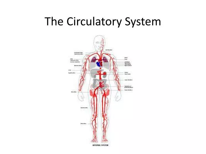
The Circulatory System. Circulatory System. The Circulatory System has two major subdivisions: The cardiovascular system: The heart The lymphatic system: Pumpless system of vessels and lymphoid organs that aids the cardiovascular system . Cardiovascular System. Location and Size.

E N D
Circulatory System • The Circulatory System has two major subdivisions: • The cardiovascular system: The heart • The lymphatic system: Pumpless system of vessels and lymphoid organs that aids the cardiovascular system
Location and Size • Location and size: • Approximately the size of a person’s fist, the hollow, cone-shaped heart weighs less than a pound • The heart is located within the bony thorax and is in between the lungs
Location and Size • Location : • Its more pointed apex is directly toward the left hip and rests on the diaphragm, approximately at the level of the fifth intercostal space • Its base, from which the great vessels of the body emerge, points toward the right shoulder and lies beneath the second rib
Coverings and wall • The heart is enclosed by a double sac of membrane, the pericardium: • The thin visceral pericardium, or epicardium, tightly hugs the external surface of the heart and is actually part of the heart wall • The fibrous layer, parietal pericardium, helps protect the heart and anchors it to surrounding structures, such as the diaphragm and the sternum
Coverings and wall • A slippery lubricating fluid is produced by the pericardial membranes: • This fluid allows the heart to beat easily in a relatively frictionless environment as the pericardial layers slide smoothly across each other
Pericarditis • Inflammation of the pericardium, often results in a decrease in the amount of fluid • This causes the pericardial layers to bind and stick to each other, forming painful adhesions that interfere with heart movements
Heart Wall - Myocardium • The heart walls are composed of three layers: • The outer epicardium, the myocardium, and the innermost endocardium • The myocardium consists of thick bundles of cardiac muscle twisted and whorled into ring like arrangement • It is the layer that actually contracts • It is reinforced internally by a dense, fibrous connective tissue network called the “ skeleton of the heart”
Heart Wall - Endocardium • The endocardium is a thin, glistening sheet of endothelium that lines the heart chambers • It is continuous with the linings of the blood vessels leaving and entering the heart
Lungs Body cells Our circulatory system is a double circulatory system. This means it has two parts parts. Pulmonary Circulation: the left side of the system: carries oxygenated blood away from the heart to the body, and returns deoxygenated blood back to the heart the right side of the system: carries deoxygenated blood away from the heart, to the lungs, and returns oxygenated blood back to the heart Systemic Circulation
Superior Vena Cava Aorta Left pulmonary artery right pulmonary artery Left pulmonary veins Pulmonary semilunar valve right pulmonary veins Left atrium Right atrium Aortic semilunar valve Tricuspid valve Bicuspid valve Right ventricle Interventricular septum Left ventricle Inferior Vena Cava
Valves – AV valves • The atrioventricular or AV valves are located between the atrial and ventricular chambers on each side • The AV valves prevent backflow into the atria when the ventricles contract • The left AV valve • Bicuspid (mitral) valve: consist of 2 cusps, or flaps, of endocardium • The right AV valve • Tricuspid valve: has 3 cusps • Tiny white cords, the chordae tendineae (heart strings), anchor the cusps to the walls of the ventricles
Valves – semilunar valves • The semilunar valves guards the bases of the two large arteries leaving the ventricular chambers • Thus, they are known as the pulmonary and aortic semilunar valves • Each semilunar valve has 3 cusps that fit tightly together when the valves are closed • When the ventricles are contracting and forcing blood out of the heart, the cusps are forced open and flattened against the walls of the arteries
Stage 1 Blood enters the heart through a vein known as the vena cava. The blood is low in oxygen. The first chamber it goes into is the right atrium. Stage 4 The blood returns from the lungs with lots of oxygen and re-enters the heart through the left atrium. Stage 5 The heart pumps the blood from the left atrium into the left Stage 2 The heart pumps the blood from the right atrium into the right ventricle. Stage 6 The heart then pumps the blood from the left ventricle out of the heart to the rest of the body through the aorta. Stage 3 The heart pumps the blood from the right ventricle through the pulmonary artery towards the lungs.
blood from the lungs blood from the body The heart beat begins when the heart muscles relax and blood flows into the atria. Cardiac Circulation • In reality, blood enters and exits both sides of the heart at the same time.
The valves close to stop blood flowing backwards. • The ventricles contract forcing the blood to leave the heart. • At the same time, the atria are relaxingand once again filling with blood. • The atria then contract and • the valves open to allow blood • into the ventricles.
Labeling exercise Label