Download
1 / 1
10 likes | 85 Vues
Imaging Findings associated with P araneoplastic Neurologic Syndromes. BAGGA,S , ESKEY CJ, FADUL, CE D ARTMOUTH -H ITCHCOCK M EDICAL C ENTER , L EBANON , NH. Purpose.

E N D
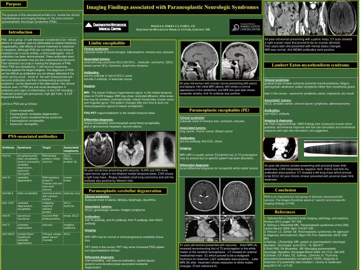
Imaging Findings associated with Paraneoplastic Neurologic Syndromes BAGGA,S, ESKEY CJ, FADUL, CEDARTMOUTH-HITCHCOCK MEDICAL CENTER, LEBANON, NH Purpose The purpose of this educational exhibit is to review the clinical manifestations and imaging findings of the most common paraneoplastic neurologic syndromes (PNS). Introduction 43-year-old woman presenting with a pelvic mass. CT scan showed a right ovarian mass that proved to be an ovarian dermoid. Four years later she presented with mental status changes. MRI was normal. Anti-MDMA antibodies were positive. • PNS are a group of rare diseases considered to be remote effects of neoplasm, and not attributable to related infections, coagulopathy, side effects of cancer treatment or extension of neoplasm. Although PNS are considered to be immune mediated only in some entities, a direct pathogenic role of antibodies has been demonstrated. These antibodies react with neuronal proteins that are also expressed by the tumor. Their detection can help in making the diagnosis of PNS. Other PNS are mediated by T cell immune responses directed against the target antigens. The diagnosis of PNS can be difficult as antibodies are not always detected & the tumor can be occult. Some of the well characterized anti-neuronal antibodies and their corresponding associated symptoms can be observed in the table below. Two clinical features seen in PNS are sub acute development of symptoms and signs of inflammation in the CSF including -- • moderate lymphocytic pleocytosis, high IgG index & CSF oligoclonal bands. • Common PNS are as follows : • Limbic encephalitis • Paraneoplastic cerebellar degeneration • Lambert Eaton myastheniforme syndrome • Paraneoplastic encephalitis • Autonomic neuropathy Limbic encephalitis Clinical syndrome: subacute onset of mood changes, hallucinations, memory loss, seizures Associated tumors: small cell lung carcinoma SCLC(40-50%) , testicular carcinoma (20%) breast cancer (8%), thymoma, lymphoma, teratoma Antibodies: anti-Hu antibody in half of SCLC cases anti-Ma-2 antibody in testicular cancer Imaging: MRI: The classic finding is hyperintense signal in the medial temporal lobes on FLAIR images, DWI may show restricted diffusion, other areas that may be similarly involved include inferior frontal lobe, insular cortex and cingulate gyrus. This pattern changes little over time & does not show progression typical of herpes encephalitis. FDG-PET: hypermetabolismin the medial temporal lobes Differential diagnosis: herpes encephalitis, post-transplant acute limbic encephalitis, glial or glioneuronal neoplasm, seizure edema. Lambert Eaton myastheniform syndrome Clinical syndrome: gradual onset of lower extremity proximal muscle weakness, fatigue, iaphragmatic weakness; bulbar symptoms milder than myasthenia gravis later in the course – autonomic symptoms, ptosis, impotence, dry mouth Associated tumors: SCLC, prostate cancer, cervical cancer, lymphoma,adenocarcinoma Antibodies: anti-VGCC antibody Imaging & diagnosis: No CNS imaging findings. EMG findings (low compound muscle action potential, decremental response with low rate stimulation but incremental response with high rate stimulation) are suggestive. 62-year-old woman with ovarian cancer presenting with ataxia and diplopia. Her initial MRI (above, left) shows a normal appearance of the cerebellum, but MRI one year later shows cerebellar atrophy. Anti-Yoantibody was positive. Paraneoplastic encephalitis (PE) Clinical syndrome: subacute onset of memory loss, confusion, seizures Associated tumors: lung cancer, Thymic cancer, Breast cancer Antibodies: anti-Hu antibody, Anti-CV2, others Imaging: MRI: MRI is usually normal. Extralimbic foci of T2 prolongation may be present but no specific pattern has been described. Differential diagnosis: broad differential diagnosis for nonspecific white matter lesions PNS-associated antibodies 62-year-old chronic smoker presenting with proximal lower limb weakness. CXR interpreted as negative. Anti- VGCC and Anti-Hu antibodies were positive. CT showed a left lung mass which proved to be SCLC62 year chronic smoker presented with proximal lower limb weakness. 56-year-old woman presenting with seizures. FLAIR and DWI show hyperintense signal in the bilateral medial temporal lobes. CXR shows a right lung mass. Biopsy revealed small lung carcinoma and anti-Hu antibody was positive by Western blot. Conclusion Paraneoplastic cerebellar degeneration • Clinical syndrome : • Subacute onset of ataxia, diplopia, dysphagia, dysarthria. • Associated tumors: • SCLC, gynecologic cancers, Hodgkin lymphoma • Antibodies: • anti-Yoantibody, anti-Hu antibody, Anti-Trantibody, Anti-VGCC antibody • Imaging: • MRI: MRI may be normal or show progressive cerebellar tissue loss. • PET: Early in the course, PET may show increased FDG uptake but hypometabolism follows. • Differential diagnosis: • viral cerebellitis, anti-seizure medication, alcohol abuse, glutamic-acid-decarboxylase-associated cerebellar degeneration PNS is an important but rare group of diseases associated with cancers. The imager should be aware of specific and nonspecific imaging findings of PNS. References 1. Osborne AG in Osborne’s brain imaging, pathology, and anatomy, Amirsys 2013, pages 767-769 2.Dalmau J, Rosenfeld MR,: Paraneoplastic syndromes of the CNS, Lancet Neurol. 2008, April ;7(4)327-340. 3. Pelosof LC, Gerber DE: Paraneoplastic syndromes; An approach to diagnosis and treatment; Mayo ClinProc September ;85(9): 838-854 4.Dalmau, J,Rosenfeld, MR: update on paraneoplastic neurologic disorders ; Oncologist :June 2010, ;15, 603-617 5.Smitt PAS, De Beukeelar JW: Managing paraneoplastic neurologic disorders: Oncologist March 2006: Vol 11(3), 292-305. 6.Erkmen, CP, Fadul, CE, Dalmau, J,Erkmen, K: Thymoma associated paraneoplastic encephalitis (TAPE): diagnosis & treatment of a potentially fatal condition. J thorac & cardiovasc surg;2011;141, e17-20. • 61-year-old woman presented with seizures. Initial MRI (A) revealed nonenhancing foci of T2 prolongation in the white matter of the cerebral hemispheres. CT showed an anterior mediastinal mass (C) which proved to be a malignant thymoma on resection. LGI1 antibodies were positive. Later MRI (B) after treatment shows resolution of white matter changes. (From reference 6)