Download
1 / 23
230 likes | 391 Vues
Introduction Structure & Support The Skeletal, Muscular and Integumentary Systems. Human Anatomy & Physiology. "Levels of Organization". Campbell/Reece Chapter 40.2. Cell Tissue Organ System entire organism. Body Tissues:. 1. Muscle Tissue function in movement, etc. Ex:

E N D
Introduction Structure & Support The Skeletal, Muscular and Integumentary Systems Human Anatomy & Physiology
"Levels of Organization" Campbell/Reece Chapter 40.2 • Cell • Tissue • Organ • System • entire organism
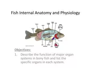
Body Tissues: 1. Muscle Tissue • function in movement, etc. • Ex: 2. Nervous Tissue: • function in communication and coordination • Ex: 3. Epithelial Tissue: • function in secretion/absorption of materials; • covering of internal/external body surfaces • Ex: 4. Connective Tissue: • functions to bind together and support other structures; forms bone and organ walls • Ex:
Humans have Bilateral Symmetry • "cephalization“ • From Latin and Greek origins: cephalicus and kephalikos respectively, both meaning "head". • anterior: • posterior: • dorsal: • ventral:
BODY CAVITIES • Cranial Cavity • Thoracic Cavity • Abdominal Cavity
BONES - The Human Skeletal System Campbell/Reece Chapter 49.5
BONE as a tissue: • strongest and hardest of all tissues • living bone continually grows and changes • "osteocyte"-bone cell Formation of bone: • Most bone begins as soft tissue (cartilage) and develops layers of bone cells • --->Ossification <---
BONE STRUCTURE: • Periosteum- tough, fibrous outer membrane, outermost covering of bone • Compact Bone- hard tube of bone tissue • Spongy Bone- softer, porous bone found at ends of bone (knobs) • Haversian Canals - network of channels through bone; contain blood vessels (makes bone porous) • Red Marrow - found inside long bones - produces, contains immature red blood cells • Yellow Marrow - found in some bones; consists of fatty materials; will produce blood cells
Cartilage Tough, flexible tissue • structural: nose, ears • protection: ends of bones (joints) • 1. cushions bones (shock absorbency) • 2. prevents bones from rubbing against each other • 3. provides smooth gliding surface • connects ribs to sternum • cartilage disks between vertebrae of backbone
The SKELETON-text reference pg. 909 Fig. 46-3 • 2 regions: • AXIAL • APPENDICULAR • Girdles: pectoral & pelvic
JOINTS • Where two bones meet Types/motion/locations: (link) • Fixed joints • Ball & Socket • Hinge • Saddle (ex: base of thumb; for rotation) • Gliding (allow bones to slide over one another; ex foot flexion) • Tendons: hold muscles to bones • Ligaments: hold adjacent bones together; in joints
DISEASES of the bone: • Osteoporosis • Arthritis
MUSCLES -The Human Muscular System • text reference: Campbell/Reece Chapter 49.6 • >7,600 muscles in human body • make up about 40% of the body’s weight • Types of Muscles • Specialized for contraction • May be: • voluntary • involuntary
"How do muscles know to move?" • Muscles are connected to nerves ("motor unit"); receive impulses from brain • Motor neurons (nervous system) control muscle contraction • 1. axon • 2. synaptic junction • 3. muscle • 4. myofibril http://highered.mcgraw-hill.com/olc/dl/120107/bio_c.swf
3 TYPES OF MUSCLE TISSUE: 1. Skeletal Muscle (A.K.A. "striated muscle") • attached to bones (connected to bones with tendons) • mostly voluntary easily fatiguable • microscopic analysis: striated (banded), with many small nuclei: Skeletal Muscle 2. Smooth Muscle (visceral) • found in organ walls (often in a circular pattern) • involuntary; ex. breathing, digestion, blood vessel diameter • fatigues slowly • microscopic analysis: flat, thin appearances, spindle-shaped, nucleated at center • for: stomach (churning) , intestine, diaphragm (raise/lower), vasoconstriction,/ vasodilation in walls of blood vessels 3. Cardiac Muscle • found in the heart • involuntary • infatiguable • microscopic analysis: slightly striated and swirled, with few visible nuclei
Action of Muscles • always work in "opposing pairs", "antagonistic" action (ex.biceps, triceps) • contract and relax (muscles do not expand-stretch appreciably) Animation http://www.yteach.co.uk/page.php/resources/view_all?id=connective_tissue_creatine_phosphate_free_fatty_acids_glycogen_myoglobin_synapse_muscle_relaxation_extensor_flexor_relaxed_muscle_contracted_muscle_tropomyosin_troponin_t_page_17&from=search
Composition of Muscle: • Muscle cells make up muscle fibers (myofibrils) which form bundles...like cables • Bundles of these muscle fibers, often hundreds of thousands, are held together by connective tissue (sarcolema) • Myofibrils are stringy, made of two types of protein: Actin (thin) and Myosin (thick) • A sarcomere is the basic unit of a muscle's cross-striated myofibril. Sarcomeres are multi-protein complexes composed of actin and myosin filaments. • The sarcomere is the basic contractile unit of a muscle (see Fig 46-12 pp. 918)
When the muscle is stimulated, the thin (actin) filaments slide past the thick (myosin) filaments. • Since the thin filaments are anchored (at the z-line), this causes the sarcomere to shorten (contract) • 1) Actin - composed of many globular actin molecules assembled in a chain. Each filament is two chains wrapped around each other • 2) Myosin- composed of bundles of myosin molecules, composed of 2 long protein chains (1,800 Amino Acids), with a globular "head" at one end • "Heads" catch in actin fibers, forming temporary "cross bridges" contraction. • animation of sarcomere shortening in action
Initiation of Contraction : The Neuromuscular Junction • Before a muscle can contract, it must be stimulated by a nerve impulse. These electrochemical signals travel across a junction (synapse) via acetylcholine - causes Ca2+ ion release = contraction • "The Motor Unit" - the axon of a single motor neuron and all of the muscle fibers that it enervates • "All or Nothing Response" • (Threshold Stimulus) • animation of neuromuscular junction at work
Regulation of Muscle Contraction • The contraction of sarcomeres is dependent upon ATP (energy) • tropomysin: regulatory protein that blocks myosin-actin attachment while muscle is at rest • When Ca2+ attaches to the troponin complex, it unblocks these binding sites, allowing the thick/thin filaments to bind, attach and cause contraction. • ATP (energy) is expended
INTEGUMENTtext reference:Ch 46-4 • The outer body covering; includes skin, hair, nails Functions of Skin: • 1st line of defense (microorganisms, disease) • helps to retain body fluids • temperature regulation • elimination of waste (sweat) • warmth
Layers: • outermost: Epidermis (dead epithelial cells) • underneath: Dermis (contains nerves, blood vessels, lymph vessels) • under dermis = fat layer (insulation) • muscle layer Glands: • sweat (cooling) • sebaceous (oil) Blood vessels: capillaries (dilate to bring blood to surface to release body heat) • Hair (follicles): for warmth • Nerves: there are receptors for pressure, touch, pain, and temperature in the skin. for explanation, try this link.
Keratin: is an extremely strong protein which is a major component in skin, hair, nails, hooves, horns, and teeth. • The amino acids which combine to form keratin have several unique properties, and depending on the levels of the various amino acids, keratin can be inflexible and hard, like hooves, or soft, as is the case with skin. • Most of the keratin that people interact with is actually dead; hair, skin, and nails are all formed from dead cells which the body sheds as new cells push up from underneath. Melanin: Also called pigment, melanin is a substance that gives the skin and hair its natural color. • It also gives color to the iris of the eye, feathers, and scales. • In humans, those with darker skin have higher amounts of melanin. By contrast, those with less pigment have lighter or more fair skin coloring. • Melanin, sometimes referred to as a chemical, is formed as part of the process of metabolizing an amino acid called tyrosine. In the skin, melanin is formed by cells called melanocytes. • more melanin is produced as a response to (and as a protection from ) UV light. • How Stuff Works: self-tanning products?