Download
1 / 24
240 likes | 506 Vues
Single-crystal X-ray diffraction at HPCAT D. Popov HPCAT Geophysical Laboratory Carnegie Institution of Washington. Why High-pressure Single Crystal Diffraction?. Reliable structure determination M ore precise refinement of crystal structures comparing to powder data

E N D
Single-crystal X-ray diffraction at HPCAT D. Popov HPCAT Geophysical Laboratory Carnegie Institution of Washington
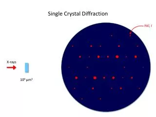
Why High-pressure Single Crystal Diffraction? • Reliable structure determination • More precise refinement of crystal structures comparing to powder data • Electron density distribution • Good use of beamtime at Bending Magnet • It is timely, because the high pressure techniques recently become available • Sufficient Q-coverage and completeness (x-ray energy, cell opening). • Matched beamsize to the sample size • Potentials for time resolved studies
HP single-crystal X-ray diffraction requirements • High energy of X-ray: typically >̴30keV • Small x-ray beam • Sufficiently large opening of high pressure cells: more than 60 • Hydrostatic (or close to) pressure medium • Precise sample controls and rotation • Intensity correction procedures • Detector with high dynamic range
HPCAT single-crystal diffraction setup Mar IP Beamstop On-line Ruby system pinhole Sample in gearbox
Temperature can be varied! Low temperature > 10K High temperature < 1000K Resistive heating On-line Ruby cryostat pinhole
Laue technique is widely used in materials science but rather new in high pressure • Mechanism of phase transitions and transformations • Deformation mechanism • Advantages with respect to application of monochromatic beam • Much faster. • Data collection in time resolved mode is possible. • There is no need to rotate the sample. • No sphere of confusion problem. • Smaller irradiated volume. • Easier to center specific area on a rotation axis. • Much more sensitive to crystal lattice deformation and rotations • Getting d-values • Exchangeable monochromators. • Absorption edges.
HPCAT Laue setup forward geometry channel cut mono detector cover photo diode pinhole CCD sample Lead pieces x/y stage • Resistive heating system • On-line Ruby • Cryostat • Detectors: • MARCCD is used routinely, • test measurements with Perkin Elmer
90 geometry CCD pinhole DAC photo diode
Commissioning test • Modeling of dislocations arrangements • Modeling of elastic strain gradients
Bent Si wafer X-ray
Bent Si wafer Beam profile 6 7 X ~(110) Z (001) 4 X-ray beam 5 20.5 4 6 2 t Deformation Ɛxx=t/R Ɛzz=ν(t/R)/(ν-1) ν=0.279 1 3 7 R=3.86mm Point 2: a= 5.433 b= 5.434 c= 5.424 γ=89.889 Point 3: a= 5.423 b= 5.422 c= 5.432 γ=90.111 15µm 5 tail tail
Streaks modeling (3-10) (1-31) 7 6 50% of peak intensity 6 (53-1) 7 7 6 (-531) 7 6 (010) (-551) (130) 6 6 7 7 6,7 oversaturated
Tails modeling (3-10) 5 (1-31) 4 3 1 10% of peak intensity 2 4 2 3 1 (53-1) 5 5 1 3 4 2 (-531) 5 3 1 2 4 (-551) (130) (010) 4 4 1 1 2 2 4,5 3 5 3 5
White Laue Technique at HPCAT • Laue diffraction setup is currently available. • Case studies using this setup are in progress. • The setup is intended for time resolved studies.
Summary • High pressure single-crystal X-ray diffraction is implemented routinely at HPCAT in temperature range 10K – 1000K. • Reliable structure determination • Information on geometry of chemical bonds • Electron density distribution • The White Laue technique is promising in time resolved studies of dislocation development and deformation under stress/pressure.