Download
1 / 39
410 likes | 524 Vues
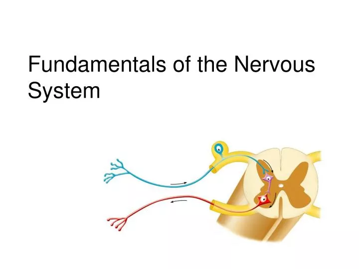
Fundamentals of the Nervous System. Basic division of the Nervous System (although there is only one NS). Central nervous system (“CNS”) – occupies cranium and vertebral column Brain Spinal cord Peripheral nervous system (“PNS”) Cranial nerves Spinal nerves

E N D
Basic division of the Nervous System (although there is only one NS) • Central nervous system (“CNS”) – occupies cranium and vertebral column • Brain • Spinal cord • Peripheral nervous system (“PNS”) • Cranial nerves • Spinal nerves • Ganglia (clusters of cell bodies)
Terminology • Input: sensory = sensory input • Receptors monitor changes • Changes called “stimuli” (sing., stimulus) • Information sent by “afferent” nerves • Integration • Info processed • Decision made about what should be done • Output: motor = motor output • Effector organs (muscles or glands) activated • Effected by “efferent” nerves Remember the difference between the English words “affect” and “effect”
Terminology, continued • “The music affected her deeply.” (Something is experienced: sensory) • “His protests had no effect.” (Something is done or not done: motor)
Nervous tissue: 2 types of cells • Neurons • Excitable nerve cells • Transmit electrical signals • Supporting cells: neuroglia or just glia • Means “nerve glue”
Neurons • All have a cell body: with nucleus and cytoplasm • Cell bodies are in clusters • CNS: clusters called nuclei • PNS: clusters are called ganglia (are outside the CNS)
Neurons, continued • Can live for a lifetime (i.e. over 100 years) • Do not divide • (exception: recent neural stem cells identified) • Cannot replace themselves • High metabolic rate • Require continuous oxygen and glucose • Die within a few minutes without oxygen
Neuron “processes” (armlike; extend from the cell body) • Nerve fibers = axons • Nerve impulse generators & transmitters • One per neuron, although can branch into “collaterals” • At terminal end branch a lot (e.g. 10,000/terminus) • Receptive regions called dendrites • Have receptors for neurotransmitters (chemicals released by other neurons) • Neurons may have many
Neuron processes • Run through CNS in tracts of white matter • Run through the PNS forming peripheral nerves
Synapses • Junctions between neurons • Information is passed (usually chemically) • Unidirectional • Presynaptic (toward synapse) vs postsynaptic (away from synapse): most neurons function as both • Synaptic cleft (tiny gap)
simplified • Info passed between neurons by chemicals • Can be excitatory or inhibitory • Along the axons, the information passes electrically
Neurons can synapse with: • Neurons • Muscle • Glands
Neurons by function/direction(relative to the CNS) • Sensory or afferent (toward CNS from sensory receptor in PNS) • Dendrites with specialized sensory receptors (in skin, muscles, viscera, etc) • Cell bodies always in ganglion* outside CNS • Motor or efferent • From CNS to muscles, glands or viscera • Cell bodies almost always in CNS* • Interneurons*: 99.98% of neurons (within CNS; can be long, e.g. travel down the spinal cord) * * *
From earlier… Nervous tissue: 2 types of cells • (Neurons and their processes: we just did) • Supporting cells = neuroglia (“nerve glue”) or just glial cells • CNS • Astrocytes • Oligodendrocytes • Microglia • Ependymal cells • PNS • Schwann cells • Satellite cells
Supporting cells • Neuroglia usually refers to CNS ones • Just “glia” to both • Divide throughout life • Smaller and darker than neurons • Outnumber neurons 10 to 1
Neuroglia (CNS glial cells) • Astrocytes • Star shaped; the most numerous • Involved in metabolism & synapse formation • Microglia • Phagocytes • Ependymal cells • Line the cavities of CNS and spinal cord; cilia • Oligodendrocytes • Produce myelin sheaths in CNS (see later slide)
PNS supporting cells • Satellite cells • Surround neuron cell body • Schwann cells • Form myelin (see next slide) in PNS
Myelin • Lipoprotein • Increases speed of conduction, large axons • Are “insulation” • Prevent leakage of electric current • Layers with spaces (nodes of Ranvier) between cells • Impulse “jumps” from node to node • “Unmyelinated” axons – smaller, slower
Myelin in the Peripheral and Central Nervous Systems In multiple sclerosis (MS), patches of myelin are destroyed in the brain and spinal cord
Schwann cells • Myelin sheath • Neurolemma (nucleus and most of cytoplasm squeezed to outside)
Gray and White Matter of the CNS(GROSS ANATOMY OF THE CNS) • Gray matter: gray-colored • Where neuron cell bodies are clustered • White matter: white-colored • Where millions of axons are running between different part of CNS, in bundles of “tracts” • Remember, tracts are in CNS, vs nerves in PNS • White is from the myelin sheaths
Usual pattern of gray/white in CNS __________________ • White exterior to gray • Gray surrounds hollow central cavity • Two regions with additional gray called “cortex” • Cerebrum: “cerebral cortex” • Cerebellum: “cerebellar cortex” ______________________________ ________________________________ (pic from Marieb lab book p 263)
Gray/White in spinal cord • Hollow central cavity (“central canal”) • Gray matter surrounds cavity • White matter surrounds gray matter (white: ascending and descending tracts of axons) • “H” shaped on cross section • Dorsal half of “H”: cell bodies of interneurons • Ventral half of “H”: cell bodies ofmotor neurons • No cortex Same pattern Dorsal (posterior) white gray Central canal_____ Ventral (anterior)
From earlier: neuron processes • Run through CNS in tracts of white matter • Run through the PNS forming peripheral nerves
Nerves are bundles of nerve fibers (long axons) in connective tissue • To or from CNS to periphery • Classified according to direction, like neurons • Mixed: carry both sensory (afferent) and motor (efferent) fibers • All spinal nerves are mixed • Sensory or afferent nerves: to CNS • Motor or efferent nerves: ventral roots of spinal cord
Interneurons(99% of all neurons) • In gray matter: • They process received sensory information • They direct this info to specific regions of the CNS • They initiate the appropriate motor response • Via axons in white matter • They transmit info (sensory and motor) from one region of the CNS to another The structural link between the PNS and CNS occurs in the gray matter of the CNS The simplest example of neuronal integration is the reflex arc (see next slide)
Reflex arcs: our “reflexes” • Fast, automatic, involuntary • Somatic or visceral • Motor responses to stimuli • Monosynaptic or polysynaptic • 5 components: see right Example of simplest, monosynapatic reflex
Basic neuronal organization • Coronal section of cerebrum • Cross sections of spinal cord and brains stem • Note gray matter (brown) and white matter (tan) • Reflex arc and information processing are shown Anterior view
Terminology for quiz • Neuron = nerve cell • Neuroglia = supporting cell • Nerve fiber = long axon • Nerve = collection of nerve fibers (axons) in PNS • Tract = collections of nerve fibers (axons) in CNS • Nucleus = cluster of cell bodies in CNS • Ganglia = cluster of cell bodies in PNS New: • Unilateral: on one side • Ipsilateral: on the same side • Contralateral: on the opposite side Remember also: • CNS vs PNS • Input: sensory: afferent: to brain • Output: motor : efferent: from brain
Pyramidal cells of cerebral cortex • This is where the “pyramidal” tract gets its name (the main motor tract from the cerebral cortex); also pyramids of medulla, pyramidal decussation