Download
1 / 50
510 likes | 712 Vues
RENAL FAILURE. DR..M.H.MUMTAZ. TYPES. 1, REVERSIBLE DYSFUNTION (acute R.failure) 2, IRREVERSIBLE DYSFUNTION (Chronic R failure). ACUTE RENAL FAILURE. 1, PRE RENAL 2, RENAL

E N D
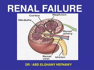
RENAL FAILURE DR..M.H.MUMTAZ
TYPES 1, REVERSIBLE DYSFUNTION (acute R.failure) 2, IRREVERSIBLE DYSFUNTION (Chronic R failure)
ACUTE RENAL FAILURE • 1, PRE RENAL • 2, RENAL • 3, POST RENAL
PRE RENAL FAILURE • CAUSES a,total body water depletion b,water redistribution ivs--------iss vasodilation,sepsis,anaphy. c,low CO--------low BP (S,M.D)
RENAL a, Interstitial nephritis b, A.T.N. hypoperfusion chemical trauma , toxins sepsis
PATHOLOGY T.obstruction T.damage T.backleakage
DIAGNOSIS a,History oligurea,concentrated U b,Tests lab. Serum,urine radiodiagnostics C.T. MRI. Ultrasount
ALTERNATIVE CLASS. Filteration failure Tubular dysfuntion Oliguric/non oliguric
Acute diseases sepsis SIRS jaundice I.A.P. renal trauma transfusion DIC Anaphylaxis muscle injury thermal burn electrocution RISK FACTORS
RISK FACTORS CHRONIC DISEASES advancing age diabetes mellitis renal disease vascular disease hyperuricaemia
Physiological changes 1. ^ age 2. ^ HR hypotension ^ CVP, lowRVPP high or low co,svr abnormal OER olig/polyurea 3. Fluid balance Oedaema high/low protein intake RISK FACTORS
RISK FACTORS Chronic drug therapy NSAIDS Diuretics Cyclosporins
Acute drug therapy A. ATN aminoglycosides cephalosporins diuretics contra. rifampicin lithium cisplatin B. Interstitial nephritis cephalosporins diuretics aspirin,NSAIDS cemetidine captopril RISK FACTORS
RISK FACTORS Proceedures a. Aortic/renal cross clamping b.Transfusion c. Major surgery
RISK FACTORS IMPAIRED RBF hypotension/m.hypertension renal art. Occlosion hepatorenal failure endotoxaemia renal vein thrombosis renal venous hypertansion
Metaboic causes 1. Electrilytes hyper-cal hypo-k hyper-phosphate 2. High oncotic P. 3. Metabolites Pigments bilirubin myoglobin haemoglobin RISK FACTORS
Post-renal urethral/blader obs. bil.ureter obs. stones/clot/tumur papillary necrosis Retroperitoneal fibrosis Surgical ligation Blader rupture Renal pelvic trauma Urethral trauma RISK FACTORS
ACUTE TUBULAR NECROSIS PHASES a,Initiation phase b,Maintenance phase
ISCHAEMIA ^ symp.stimulation ^ renin activity PGE2 ANH inhibition ^ ADH ^ adenosine ^ endothelin NEPHROTOXINS Ischaemia increases the susceptibility to nephrotoxic agents INITIATION PHASE
MANTENANCE PHASE • Factors acting to maintain filteration failure 1,tubular obstruction 2,tubular backleak 3,vasodilatation of efferent art. 4,decreased GMP
Mechanism of oligurea a,glomerulo-tubular balance b,decreased GMP c,itratubular obstruction d,interstitial oedema e,cortical ischaemia
A,oligurea absolute relative B, azotaemia normal solute load maximum in catabolic states in ARF ^ urea/d ^ cr/d Complications of ARF/ATN
C,Biochamical ^NaCl/water ^ K ^ HPO4 hypocalcaemia ^ Mg ^ uric acid M.acidosis D,Haematological Anaemia Thrombocytopaenia Leukocyte dysf. Complications
E,Immunosupression Lumphopaenia Reduced IgG Reduced comple. Impaired PMN R.I.response Drug effects Infections F,C.V.S. CCF Hypertention Arrhythmias Pericarditis Effusion Complications
G, G.I.T. Anorexia,Nausea, Ileus,Hmge. H,Neurological Lethargy,somnolance Confusion, Convulsions ^ sensitivity to anaesthetics Complications
Complications I,causes of pulmonary infilterates in ARF 1,LVF/CCF 2,bacterial pmeumonia 3,Atypical pneumonia 4,Septicaemia 5,ARDS 6,Autoammune diseases
A,Tubular dysfuntion B,Glomerular dysfuntion C,Other causes low C.O. Resp.F Starvation Rhabdomyolysis Hyperkalaemia Organic acids Causes of Acidosis in ARF
Biochemistry INVESTIGATIONS IN ARF
Definitions • RFI=RENA FAILURE INDEX • =urine(Na)/(U/P creatinine) • FEna=%fractional excretio Na • =(U/P Na).100/(U/P creatinine)
Abnormal urea/creatinine ratio • Normal U:C ratio 100:1( R;70-150) • Pre-renal disease >200:1
Abnormal urea/creatinine ratio • High Ratio • ^ urea .dehydration/hypovol. • .GIT.bleeding • .Catabolic state • .Hyperalimentation • .Drugs • low creatinie .elderly,low m. mass
Abnormal urea/creatinine ratio • Low Ratio • low urea. Liver failure • hepato-renal synd • Malnutrition • High creatinie rhabdomyolysis • acute m.disease • ketones,drugs
CREATININE CLEARANCE • 1, clearance(ml/min=(N-age[years])*BW(kg)/serum creat. N = 150 foe female N = 160 for male > 70 N = 170 for male < 70 2, clearance(ml/min)=UV*1000 /p*420 U=urine creatinine level V=urine volume (midnight &7 am) P= plasma creatinine level
2. Urinary sediment • .Cast types • i,hyaline casts, fever,diuretics,RD • ii,red cell casts glomerulonephritis • iii,w.cell casts pyelonephritis • iv,waxy casts chronic renal disease
3,Imaging • 1, Ultrasound • 2, CT scan • 3, IV pylogram • 4, radio-isotope perfusion scan • 5, renal angiogram
4,Renal biopsy • 1, glomerulonephritis • 2, vasculitis • 3, SLE • 4, Goodpasture syndrome • 5, TTP • 6, Interstitial nephritis • 7, oligurea lasting > 8 weeks
Renal failureprophylaxis&protection • Methods • 1, physiological • 2,physical • 3,pharmacological • 4,replacement therapies
Physiological methods • a, normalise blood volume • iv fluids,(Na containg) • b,optimise cardiac output • iv fluids.inotropes,vasopressors • c, optimise O2 delivery • Hb,Spo2,avoid acidosis • d, high sodium excretion
Physical methods • Detection/management of IOH • Detection/management-post renal obs. • Limitation of aortic clamp times • Avoidance of embolisation • Minimise direct trauma
Pharmacological methods • Avoid nephrotoxins • Avoid inhibitors of autoregulation • Diuretics • Renodilators • Other agents • free radical scavengers • Ca channel blockers
Renal replacement methods • Haemo- filtration • Haemo-diafiltration • Haemodialysis • R. Transplant.
Renal failure---Frusemide • Beneficial effects • Increased tubular&urine flow • Increase Na &osmolar clearance • Decreased tubular O2 demand • Stimulate vasodilator prostaglandins • Deleterious effects • Hypovolaemia • Hypokalamia,Hyponatraemia • Ototoxicity
Uses in non renal failure • Fluid overload • Cerebral oedema • Hyperkalaemia • Renal protection • ( decreased O2 demand)
Renal failure---Mannitol • 1,Osmotic diuresis • 2,Anti sludging ,tubular protect. • 3,renal vasodilatory PG synthesis • 4,Free radical scavenger • 5,Decreased T. swelling
Renal failure---Dopamine • Increases Fe Na excretion • Increases urine out put • Does not increase creatinine clearance • Inotropic effect • Doesnot prevent ac.renal failure • Side effects, • gastric stasis,inhibition of • ant pit.hormones,hypoxic • drive depression.
Renal failure---Nor-adrenaline • Increases perfusion pressure by increase • of efferent arteriolar resistance • more than afferent art.resistance
Other therapies • 1,Calcium channel blockers • 2,Adenicine recepter antagonists • 3,Oxypentifylline • 4,Chlorpromazine • 5,Clonidine • 6,ATP-MgCl2 • 7,ANF
Conclusion,Renal rescue therapy • Normalise;- • Blood flow • blood volume • blood pressure • O2 delivery • CO—CI • Blood Pressure, s,m,d.