Download
1 / 22
230 likes | 519 Vues
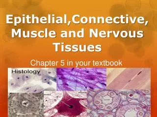
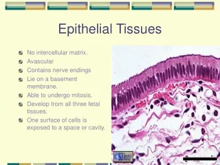
Histology Slides for the Epithelial, Connective, and Integumentary Tissues. Slides are presented in order of magnification As you view the following slides make sure you can accomplish these goals: Can you identify the tissue layers observable on the slides?

E N D
Histology Slides for the Epithelial, Connective, and Integumentary Tissues • Slides are presented in order of magnification • As you view the following slides make sure you can accomplish these goals: • Can you identify the tissue layers observable on the slides? • Can you identify the specific structures or layers indicated by the numbered arrows or brackets? • At the end of a sequence you will find the answers to the above for each vessel and layer.
Identify cells seen in this view? How is their shape related to function?
Identify cells seen in this view? How is their shape related to function?
Identify cells seen in this view? How is their shape related to function?
Identify cells seen in this view? How is their shape related to function?
Identify stages of Mitosis 2 4 1 5
Answers to Slides 2-8 Slide 2: Surface view of simple Squamous epithelial Cells designed to cover surfaces, have large surface area on surface plane and are thin in side view. Slide 3: Smooth muscle cells Cell spindle-shaped to contract and elongate on a single plane Slide 4: Red blood cells with a few white blood cells Biconcave to increase surface area to volume for diffusion and non-nucleated to increase hemoglobin capacity. Slide 5: Human sperm cells Small flagellated cells for swimming. Small size to increase the number of cells that can be placed in a small amount of solution Slide 6-8: Mitosis of a fish egg 1. Metaphase 4. Early telophase 2. Prophase 5. Late telophase 3. Anaphase
2 1
1 1
4 1 3
1 3
Answers to Slides 10-13 Slide 10: Kidney showing a renal corpuscle Slide 11: View of lung tissue Slide 12: Artery low power Slide 13: Artery high power 1. Side view of simple squamous tissue 2. View of simple cubiodal 3. Dense irregular Connective Tissue 4. Adipose tissue
6 2
Answers to Slides 15-21 Slide 15: View showing kidney tubules Slide 16: View of thyroid gland Slide 17: View of small intestine villus Slide 18: X-section through a bronchiole Slide 19: Esophageal epithelium Slide 20: Vaginal epithelium Slide 21: Epidermis of non-hairy skin 1. View of simple cuboidal epithelium 2. Simple columnar epithelium 3. Pseudostratified columnar epithelium 4. Nonkeratinized stratified squamous epithelium 5. Keratinized stratified squamous epithelium 6. Goblet Cell with a mucous excretory vesicle