Download
1 / 33
371 likes | 620 Vues
DNA Structure and Genetic Variation. Genetics Spring 2014. Outline. Genetic Differences Among Individuals. DNA sequences can be single-copy or repetitive and can also be clustered or interspersed . In humans and in 23 pairs of chromosomes, we found that:.

E N D
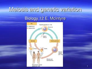
DNA Structure and Genetic Variation Genetics Spring 2014
Genetic Differences Among Individuals DNA sequences can be single-copy or repetitive and can also be clustered or interspersed. In humans and in 23 pairs of chromosomes, we found that: • 1.5 % of genome encodes polypeptides • 5% of genome contains regulatory sequences • 50% of the genome contains unique DNA sequences • 50% of the genome contains repetitive DNA sequences • 99.9% of genome is shared among all humans LINEs: Long INterspersed Elements SINEs: Short INterspersed Elements LTRs: Long Terminal Repeats (Retrotransposons)
Pseudogenes are non-coding DNA sequences that arose from duplication or retrotransposition (evolutionary process). Most gene families have pseudogenes, they are related to genes and can revert to being expressed. Pseudogenes are non-functional genes that can be either non-processed (classical) or processed. LINES, SINES, and processed pseudogenes, are all retroposons. The retroposon process is when a DNA sequence is transcribed, making a mRNA copy. The copy is converted into DNA by reverse transcriptase then the DNA piece is inserted back into the chromosome DNA. Both b-globin and OR gene families contain numerous pseudogenes.
Molecular Structure of DNA: Monomers A deoxynucleotide contains a base, a sugar (dioxyribose), and phosphate group. Dioxynucleoside = base + sugar Dioxyucleotide = base + sugar + phosphate The RNA molecule is formed of nucleotide (base, ribose and phosphate group)
Molecular Structure of DNA: Phosphodiester Bonds Nucleotides join to form a DNA or RNA chain Polynucleotide chains have 5’-PO4- at one end and 3’-OH on the other end
Molecular Structure of DNA: Anti-parallel Structure DNA strands are anti-parallel 5’->3’ ; 3’->5’
Separation and Identification of Genomic DNA Fragments • Nucleic Acid Hybridization (Southern Blot) • To identify a specific DNA fragment in genomic DNA; need specific probe. • Polymerase Chain Reaction (PCR) • To specifically and repeatedly replicate (amplify) one fragment from genomic DNA; need 2 specific primers.
Separation and Identification of Genomic DNA Fragments: Southern Blot • Named after biologist Pr. Edwin Southern • A Southern blot can tell: • Whether a particular gene is present and how many copies are present in the genome of an organism • The degree of similarity between the chromosomal gene and the probe sequence (cannot pick up small mutations) • Whether recognition sites for particular restriction endonucleases are present in the gene • Whether re-arrangements have occurred during the cloning process • DNA fingerprinting, which is a technique used to compare two samples of DNA • Restriction map
Gel Electrophoresis Migration of DNA in a gel is related to DNA size DNA molecules must pass through pores in the gel - smaller fragments migrate more rapidly
Standard Curve • A standard curve is a plot of known units (DNA sizes of the molecular weight markers, Y axis) versus their corresponding experimental values (distance traveled after electrophoresis in this exercise, X axis) • It allows for the identification of unknown comparable units or for the confirmation of known comparable units (DNA sizes of restriction digests or PCR products). Graph of agarose concentration and size range of DNA fragments that can be separated
Applications: Genetic Disease Diagnostic Androgen insensitivity syndrome • cDNA probe for human androgen receptor gene • Genotype is 46, XY but the phenotype is female South African athlete Caster Semenya won several gold medals (results of gender testing were not released)
Applications: Ribotyping (fingerprinting of genomic DNA encoding rRNA in bacteria) Glycopeptide Resistance among Coagulase-Negative Staphylococci that Cause Bacteremia. Southern blot of chromosomal DNA showing a representative sample of the different patterns obtained by ribotyping of the analyzed Staphylococcus epidermidis isolates. Lane A, strain 559 (intensive care unit [ICU]); lane B, strain 528 (ICU); lane C, strain 520 (ICU); lane D, strain 428 (cardiosurgery); lane E, strain 427 (medicine); lane F, strain 346 (infectious diseases); lane G, strain 345 (ICU); lane H, strain 95 (medicine); lane I, strain 93 (cardiosurgery); lane L, strain 29 (ICU); and lane M, λ HindIII marker.
Separation and Identification of Genomic DNA Fragments: PCR • PCR amplifies a specific DNA fragment • PCR requires two primers, one at each end of target • Each cycle doubles the amount of target DNA • Repeated cycles produce millions or billions of copies • RT-PCR looks at mRNA expression
Types of DNA Markers in Genomic DNA and their Applications • There is about 99.9% of identity in the nuclear DNA of any two humans. • In a random 1,000 bp nuclear DNA sequence, there is only one bp that varies between two homologous chromosomes inherited from unrelated parents. • In a random 2, 500 bp coding nuclear DNA sequence, there is only one bp that varies between two homologous chromosomes inherited from unrelated parents. Selective pressure Genetic polymorphism refers to variants found in 1% or more of chromosomes. Rare variants refers to variants found in less than 1% or of chromosomes. Forensic medicine was derived from genetic polymorphism research. Personalized medicine was derived from genetic polymorphism research.
DNA Markers or DNA polymorphisms are genetic variants that appear in at least 1% of a population. A condition in which one of two different but normal nucleotide sequences can exist at a particular site in a DNA molecule.
Polymorphism: Single Nucleotide Polymorphisms (SNPs) Need DNA Sequencing Oligonucleotides attached to a glass slide in a SNP chip can be used to identify duplex DNA molecules containing alternative base pairs for a SNP, in this example a T–A base pair versus a C–G base pair. The SNP genotype of an individual can be determined by hybridization because DNA from genotypes different give a different pattern of fluorescence
The International HapMap Project is a multi-country effort to identify and catalog genetic similarities and differences in human beings. Using the information in the HapMap, researchers will be able to find genes that affect health, disease, and individual responses to medications and environmental factors. The Project is a collaboration among scientists and funding agencies from Japan, the United Kingdom, Canada, China, Nigeria, and the United States.
Polymorphism: Restriction Fragment Length Polymorphisms (RFLPs) RFLPs are SNPs located within a restriction enzyme site. Does not need DNA Sequencing A difference in the DNA sequence of two molecules can be detected if the difference eliminates a restriction site
In a restriction fragment length polymorphism (RFLP), alleles may differ in the presence or absence of a cleavage site in the DNA RFLPs are codominant • RFLPs create DNA fragments of different sizes. • The fragments are the same size in a homozygote. • The fragments are different sizes in a heterozygote.
Polymorphism: STRP, short tandem repeat polymorphism (microsatellite) and VNTR, variable number tandem repeat (minisatellite) Different numbers of repeats can be distinguished by PCR using primers to the flanking DNA or by Southern blot (later slides)
Microsatellite markers in human DNA. At top is the DNA containing an (AC)n microsatellite marker on one chromosome; primers 1 and 2 are PCR primers complementary to unique sequences that flank the dinucleotide repeat. Below is a pedigree demonstrating codominant inheritance of a microsatellite polymorphism due to variable numbers of the dinucleotide AC. The genotype of each individual is shown below his or her symbol in the pedigree. The different-sized fragments are amplified by PCR with primers 1 and 2 flanking the stretch of AC dinucleotides, and their relative lengths are determined by separating them by gel electrophoresis (bottom).
CODIS: Combined DNA Index System contains the DNA profiles contributed by federal, state, and local participating forensic laboratories. 13 CODIS Core STR loci with chromosomal positions (Amelogenin tests for sex)
F.B.I. crime lab What is Norma’s genotype?
Minisatellite: Codominant inheritance of an autosomal DNA polymorphism caused by a variable number of tandem repeats. Alleles 1 to 4 are related to one other by a variable number of identical (or nearly so) short DNA sequences (arrows). Size variation can be detected after restriction enzyme digestion and hybridization with a unique probe that lies outside the VNTR sequences themselves but inside the restriction sites used to define the allelic fragments.
Minisatellite: DNA fingerprinting of twins by means of a probe that detects VNTR polymorphisms at many loci around the genome. Each pair of lanes contains DNA from a set of twins. The twins of the first set (as well as the twins of the third set) have identical DNA fingerprints, indicating that they are identical (monozygotic) twins. The set in the middle have clearly distinguishable DNA fingerprints, indicating that they are fraternal twins.
Polymorphism: Copy-Number Polymorphism (CNP) CNPs were discovered by the application of the newer technology: array comparative genome hybridization (CNV: copy number variation). Chromosome 3 showing a loss of chromosomal material denoted by the red bar. The karyotype shows other chromosomal gains (green bars) and losses (red bars) within the genome.