Download
1 / 91
950 likes | 1.44k Vues
Parasitology Course BIOL 2272. By Fred Opperdoes. Definition of parasitism. Parasitology describes the relationships between two organisms, i.e. the host organism and the parasite. When one organism gives shelter and food to another organism two totally different situations may exist:

E N D
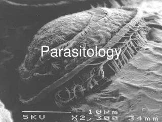
Parasitology Course BIOL 2272 By Fred Opperdoes
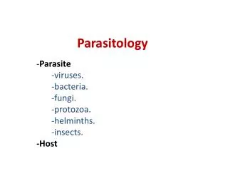
Definition of parasitism • Parasitology describes the relationships between two organisms, i.e. the host organism and the parasite. When one organism gives shelter and food to another organism two totally different situations may exist: • Without any harm being done, or in the case where some hosts may benefit from the presence of a parasite, in which case we normally speak of: • mutualism: a situation where two organisms live in some contact and benefit from each others presence. They exchange food or provide shelter or protection, but may still be able to live an independent life. • symbiosis: a situation where two organisms live in close contact and benefit from each others presence. They have become dependent upon each others presence and are unable to survive independently. • endosymbiosis: bacteria have invaded the cytoplasm of a eukaryotic host cell. Host cell and bacterium have become dependent upon each other. In the case large amounts of genetic material have been moved from the bacterial genome to the nucleus of the host an irreversible situation is reached which led to the formation of: • chloroplasts, photosynthetic organelles that have originated from photosynthetic cyanobacteria • mitochondria, organelles involved in oxidative phosphorylation that have originated from the alpha subdivision of the proteobacteria.
Definition of parasitism (cont.) • With harm being done. • When one organism lives at the expense of its host we can distinct several situations: • * viral infections • * bacterial infections • * parasitic diseases • NB: Normally a true parasite does not kill its host
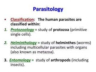
Parasitic associations are very common in Nature • There is no major taxonomic group that has no parasitism associated with it. • Only viruses have no parasites ! Host organism ParasiteViruses -Prokaryotes Eubacteria and Archaebacteria bacteriophages/plasmids Eukaryotes Protozoa viruses and bacteria Fungi viruses and plasmids Metazoan organisms viruses and bacteria, protozoa and fungi and metazoan organisms • In most cases, however, the associations presented in the above table are not regarded as true parasitism and are dealt with by such disciplines as virology, microbiology and mycology (the study of fungal infections). Only when metazoan organisms are infected by protozoans, or by helminths, or infested by organisms that live in or upon the host's skin (ectoparasites), this is regarded as a case of parasitism.
What is parasitology (1) • Parasitology is the study of parasitism and due to the nature of parasitism it can not be well defined. • Parasitology can be divided in : • The study of the parasite itself. It is important to know how the parasite functions in order to be able to combat it. This comprises all disciplines of biology and beyond. • Host-Parasite relationships • This comprises all disciplines that deal with the host-parasite interface and the way the two organisms interact with each other. The most important disciplines are: • Physiology • Biochemistry • Cell biology • Immunology • Pharmacology
What is parasitology (2) • Immunology of parasitic infections. This deals with the description of the immunological response by the host and the way the parasite tries to evade the host's immune attack. • This comprises such disciplines as: • Humural and cellular immunology • Molecular immunology • Chemotherapy of parasitic infections. This deals with the study of the effect of drugs on the parasite and host and the treatment by drugs that would lead to the killing of the parasite and the recovery of the diseased host. • The disciplines involved are: • Organic chemistry • Pharmacology • Biochemistry • Medicine • Epidemiology tries to describe the ways the diseases spread amongst the population. In the case of parasitic infections not only the host, but also the parasite and the vector should be included.The disciplines are: • Tropical hygiene • Entomology • Geography
Different types of parasites • Due to the great variety in parasitic organisms in Nature they can be divided in many different classes:Type of parasite Examples(Viruses) -(Bacteria) -Phytoparasites Yeasts (Candida),FungiZooparasitesEctoparasites Bugs, lice (punaises, poux)Endoparasites bloodvessels: Schistosoma intestin: Taenia tissues: Trichinella cells: Leishmania, Toxoplasma
Different types of parasites (2) • Parasites can be divided into different classes of parasite : • Obligatory parasites. • These parasites can only survive in a host and therefore go directly from one host to another. This may involve complex life cycles. • Examples are: • Trichomonas vaginalis • Taenia • Trichinella
Trichomonas vaginalis (trichomoniasis) • Trichomonas vaginalis is a sexually transmitted disease (STD), although transmission by other routes (such as soiled towels) has been documented. There is no cyst in the life cycle, so transmission is via the trophozoite stage only. Most people infected with trichomoniasis are asymptomatic. It is one of the most frequent sexually transmitted diseases in man. 30 to 50 % of all women in western countries may be infected. In males the infection normally does not lead to any symptoms, while in females this may lead to an increased secretion, characterized by a white discharge from the genital tract and itching. Diagnosis depends on finding trophozoites in secretions of the genital tract from men or women. In cases where the numbers of organisms are very low, the trophozoites can be cultured to increase their numbers. Light microscopic picture of Trichomonas vaginalis in culture
Trichomonas vaginalis (trichomoniasis) • Trichomonas vaginalis is a facultative anaerobic protozoan parasite that has no mitochondria or peroxisomes. Trichomoniasisis treated with metronidazole (Flagyl). The drug is reduced inside the parasite by the enzyme hydrogenase located in typical organelles called hydrogenosomes. When the drug reacts with traces of oxygen present in the organism it forms oxygen radicals that rapidly kill the parasite. Metronidazole is also used for the treatment of Giardia and Entamoeba infections and for infections by some anaerobic bacteria. A trophozoite of Trichomonas vaginalis from culture. The four flagella and single nucleus are visible. The dark median rod is the axostyle which is characteristic of the trichomonads; approximate size = 26 µm.
Different types of parasites (3) • Temporary parasites. • These parasites spend only part of their lives as a parasite and another part as free-living organism. • Examples are: • Fasciola hepatica [Liver fluke (douve)] • Schistosoma • Ascaris • Haemonchus
Different types of parasites (4) • Facultative parasites. • These organisms are normally free living and infect a host only by accident. • Examples: some free-living amoeba such as: • Naegleria • AcanthamoebaTrophozoites of Acanthamoeba castellani • Fungi such as Candida(vaginal candidosis, athlete foot)
Naegleria • The genus Naegleria includes several species of free-living uninucleate amoebae that live in aquatic environment (ponds, lakes, sewage). It belongs to a family of amoebae (Valkampfiidae) whose members can transform into flagellate stage, which is a transient, non-feeding non-dividing form; the amoebal stage can also transform into a resistant cyst stage. The species Naegleria fowleri is occasionally pathogenic to a variety of mammals including humans. Following direct intranasal contact, usually by swimming, N. fowleri invade the olfactory bulb and progress along the olfactory nerves to the brain, thereby causing an acute and fulminating disease called primary amoebic meningoencephalitis. Naegleria trophozoites in brain tissue
Acanthamoeba • Acanthamoeba spp. (Rhizopoda; hartmanellidae) is a free-living amoeba which is common in a variety of habitats, including fresh water, soil, and sewage. In humans they can cause a sub acute or chronic infection called granulomatous amoebic encephalitis. Acanthamoeba is frequently found in contact lenses solutions, thereby causing vision-threatening keratitis. Despite morphological similarities with Naegleria and other free-living amoebae, sequence comparisons arising from small and large ribosomal RNA subunits suggest Acantamoeba being as closely related to higher plants and vertebrates as they are to various protist genera. Acanthamoeba trophozoites in culture after immunofluorescence staining
Athlete foot or Tinea pedis • Athlete foot or tinea pedisis a common condition in occupational groups and communities. In boarding schools and amongst coal miners it may reach epidemic proportions. The commonest organisms are T. interdigitale and T. rubrum and the high rate of infection is associated with its increased opportunity to spread for instance in shower rooms, where the parasite dwells in the wooden 'duck' boards in the showers. Preventive measures consist in the removal of those wooden boards from the showers, regular foot inspection and the use of bathroom footwear. • Treatmentconsist in the application onto the infected skin of miconazole nitrate ointment (Daktarin, Janssen Pharmaceutica), the use of miconazole-nitrate foot powder in socks and shoes and the oral use of Sporanox (isoconazole nitrate). The mode of action of these imadazoles is based upon their capacity to inhibit the action of fungal cytochrome P450 that is involved in the biosynthesis of ergosterol, an essential sterol of the fungal plasma membrane.
Vaginal candidosis • The diseaseIn recent years there has been a dramatic rise in the incidence of vaginal candidosis. Such Candida infections have been associated with a number of factors most notably diabetis mellitus and pregnancy. The contraceptive pill and an increased sexual activity have also been held responsible. Recurrent and even chronic infections in women are common. • Treatmentconsist in the use of oral fungicides or the application of miconazole nitrate ointment (Daktarin, Janssen Pharmaceutica) into the vagina (for mode of action see athlete foot).
Types of host • Like the parasites, the hosts can be divided into several classes as well. These are: • Definitive host (DH).A definitive host is an organism that hosts the adult (sexual) form of the parasite • Intermediate host (IH).An intermediate host is an organism that hosts the asexual form of the parasite (only when there is an obligatory passage through the host). Intermediate hosts can be divided into two groups: • Passive IH (molluscs in the case of Schistosoma) • Active IH (tsetse fly in the case of trypanosomes) • Also the vectors can be divided into two different types: • Biological vectors • Examples: haematophagous athropodes such as mosquitoes and other biting insects • Mechanical vectors • Examples: flies for transport of amoebal cysts
Mechanisms of infection • A parasitic infection may occur by various routes, depending on the nature of both parasite and host • Infection by passive entry. • By feeding and drinking . Cysts and eggs can be tranported by water or by insects such as the housefly. Examples are: Giardia. Giardia, an intestinal parasite of beavers and man may contaminate fresh water lakes and rivers in the US. The drinking of this water may lead to intestinal infections in humans. The trophozoites contain a dark transverse rod, the axostyle, which seems to be a supportive element. The cysts average about 13 µm in length, are oval, and contain two nuclei and remnants of the axostyle. Because of these unique characteristics, G. lamblia is one of the easiest intestinal protozoans of humans to diagnose. Giardia trophozoites in the stool
Giardia lamblia • G. lambliatrophozoites adhere closely to the lining of the small intestine, and in heavy infections much of the lining of the small intestine can be covered with trophozoites. The symptoms associated with giardiasis range from none (in light infections) to severe, chronic diarrhea (in heavy infections), but not dysentery. • One person can pass millions of G. lamblia cysts each day, and most infections probably result from water or food contaminated with human sewage. Open sewers in city streets and contamination of drinking water with this sewage undoubtedly results in many infections. However, in some countries the use of human fecal material ("night soil") as a fertilizer is also an important source of infection. Many cases of "traveler's diarrhea" are caused by Giardia. Even in developed countries potable water can be contaminated with small amounts of sewage, especially when septic systems are build too close to wells. G. lamblia is found throughout the world. • The parasite is also transmitted through anal sexual intercourse by homosexuals males.
Mechanisms of infection (2) • Entamoeba, an intestinal parasite of humans is transmitted in the form of cysts by the house fly. • Entamoeba trophozoites in the stool
Entamoeba histolytica • Entamoeba histolytica is a facultative anaerobic protist that forms trophozoites as well as cysts. • The life cycle of Entamoeba histolytica involves trophozoites that live in the host's large intestine and cysts that are passed in the host's feces. Humans are infected by ingesting cysts, most often via food or water contaminated with human fecal material. The trophozoites can destroy the tissues that line the host's large intestine, so of the amoeba infecting the human gastrointestinal tract, E. histolytica is potentially the most pathogenic. In most infected humans the symptoms of "amoebiasis" are intermittent and mild (various gastrointestinal upsets, including colitis and diarrhea). In more severe cases the gastrointestinal tract hemorrhages, resulting in dysentery. In some cases the trophozoites will enter the circulatory system and infect other organs, most often the liver (hepatic amoebiasis), or they may penetrate the gastrointestnal tract resulting in acute peritonitis; such cases are often fatal. As with most of the amoeba, infections of E. histolytica are often diagnosed by demonstrating cysts or trophozoites in a stool sample.
Mechanisms of infection (3) • Naegleria fowleri, present in surface water, may enter via the nose when swimming and this may lead to a fatal amoebal meningoencephalitis. • Naegleria fowleri in brain tissue
Mechanisms of infection (4) • Acanthamoeba, a facultative parasite may contaminate soft contact lenses and cause eye infections. • Acanthamoeba trophozoites in culture
Mechanisms of infection (5) • By direct contact such as sexual intercourse • Trichomonas, is transmitted via sexual intercourse and is the causative agent of the most common sexually transmitted disease amongst humans • Giardiais widespread amongst the San Francisco gay community. • Trypanosoma equinum is a parasite of equines (horses) and is transmitted via sexual intercourse • * Trichomonas vaginalis trophozoites Trypanosomes in blood Giardia lamblia trophozoites in the stool
Mechanisms of infection (5) • Active entry • By biting (haematophagous) insects that serve as the active vectors in the transmission of disease: • mosquitoes transmit malaria and filariasis • black flies transmit onchocerciasis or river blindness • tsetse flies transmit sleeping sickness in Africa • sandflies transmit leishmaniasis • By highly specialized developmental forms in the life cycle of the parasite • Schistosoma cercaria actively penetrate human skin Anopheles mosquito Tsetse fly sandfly fly
Parasite reservoirs • A parasite reservoir (PR) is the biotope where the parasite lives. In general it is the Definitive Host, where lives the sexual stage of the parasite, producing large amounts of larvae or eggs, that serves as the PR. A PR can be: • Human reservoir • Human is the sole host (schistosomiasis) • Human is an accidental host (leishmaniasis) • Animal reservoir • 1. domestic animals • 2. wild animals
How do parasites survive inside an immunocompetent host ?(1) • Ectoparasites • Infestation leads to local lesions of minor to moderate importance. This leads only to: allergic reactions (itching) and immunological reactions (without result) • None of these reactions really harm the parasite, but do harm the host • Intracellular parasites • These parasites try to escape any immunological reactions mounted by the host by hiding themselves inside the host cells where the immune system cannot reach them. • Examples are: • Toxoplasma in lymphocytes, • Plasmodium in erythrocytes, • Leishmania, in macrophages, • T.cruzi in muscle cells.
How do parasites survive inside an immunocompetent host ?(2) • Extracellular parasites • Some parasites cover their cell surface with host serum proteins to avoid recognition by the immune system of the host • Examples are: • Schistosoma worms that cover themselves with host serum albumin • Rodent trypanosomes that cover themselves with ablastin (IgE) • Antigenic variation in the African trypanosomes that live freely in the bloodstream and body fluids of the host is another effective mechanism of evasion. • Cyst formation by Entamoeba and other amoeboid parasites
Harmful effects of the parasites on the host (1) • Many parasites cause harmful effects to their host, but in most cases these effects are not of such importance that the host is being killed. • Such effects comprise: • Wasting (cachexia, spoliatrices)African trypanosomiasis and leishmaniasis may lead to severe loss of weight in both animals and man. • Superinfections In the case of (muco)cutaneous leishmaniasis ulcerations may lead to superinfections with bacteria • Production of toxic compoundsIt is thought that the African trypanosome, when in the central nervous system, produces aromatic amino-acid analogues that may influence brain function. • ImmunodepressionMalaria, bilharziosis, etc., lead to a certain degree of immune suppression which renders the infected host more susceptible to other diseases. • Allergic reactionsIn the case of onchocerciasis (river blindness) the presence of the filarial worms under the skin may lead to depigmentation due to allergic reactions.
Harmful effects of the parasites on the host (2) • Anaphylactic shock • may be induced by the sudden release of large amounts of parasite internal antigens into the bloodstream. • In malaria this occurs when the merozoites are released in waves from infected erythrocytes. • In African trypanosomiasis or sleeping sickness this occurs when the immune response leads to the massive killing and lysis of the circulating parasites. • Also drug treatment leading to a massive killing of the parasites may result in anaphylactic shock. • Mechanical damage • In the case of malaria the lysis of erythrocytes does lead to haemolysis and anaemia. • In the case of ascaris infection the presence of the worms in the small intestine may lead to intestinal occlusions • Irritative reflexes (intestinal contractions: ascaris) • Irritation of skin and tissues by ecto- and endoparasites
Prophylaxis (1) • Prophylaxis is regarded the prevention of disease by taking special measures. These measures may comprise a great variety of individual as well as collective measures • Prophylactic measures and their examples are: • Isolate the parasite reservoir (PR)In the case of a zoonozis such as leishmaniasis it would be sufficient to remove the infected dogs from the houshold. • Sterilize PR In the case of schistosomiasis the PR can be sterilized either by treatment of the population by chemotherapy with drugs or by hygienic measures such as the placement of latrines in or just outside the villages. • Eliminate intermediate hostIn the case of schistosomiasis treatment of lakes and rivers with molluscicides would interupt transmission via the snail as intermediate host.
Prophylaxis (2) • Eliminate vectorsTreatment of surface water with larvicides or the application of insecticides in the house in the case of malaria will reduce or prevent transmission. • Isolate vectorsThe use of bed nets, impregnated or not, with insecticides is an effective protection against mosquito bites. • VaccinateThis would be the most efficient protective measure. However, so far there is no vaccine available for any parasitic disease of humans. • Carry out chemoprophylaxisThe preventive use of chloroquine (Nivaquine) for long periods when travelling in areas where malaria is endemic • Depending of the nature of the parasite, its host and its vector different measures or a combination of measures have to be taken.
The Kinetoplastida • Trypanosomiases and leishmaniases are caused by haemoflagellated protozoan parasites that belong to the family of the Trypanosomatidae. • Trypanosomatidae, or trypanosomes, are members of the protist order Kinetoplastida, which can be divided in Bodonina and Trypanosomatina. Contrary to the Bodonina, which can be divided in the free-living Bodonidae and the parasitic Cryptobiae, all Trypanosomatidae, without exception, are parasitic. They are flagellated and parasitise all major groups of multicellular organisms such as insects, plants and animals. They can either be extracellular or intracellular and have a great variety of shapes, depending on the stage of the life cycle and their location within the host.
The Kinetoplastida • The following parasite-host associations are known: • Trypanosomatidae • Insect parasites • Crithidia • Herpetomonas • Plant parasites • Phytomonas • Amphibian parasites • Trypanosoma mega • Fish parasites • Trypanosoma • Reptiles • Leptomonas • Animals • Trypanosoma • Leishmania • Cryptobiae • Fishes • Trypanoplasma • Bodonidae • free-living
Morphology of trypanosomes Scanning EM of blood from an experimental animal (rat) infected with Trypanosoma rhodesiense with a very high parasitaemia (2-3 billion tryps / ml). There are almost as many trypanosomes as there are red blood cells. Diagrammatic representation of a short stumpy bloodstream form trypanosome (from K. Vickerman with permission). A-the anterior flagellum. B-the underlying complex cytoskeleton. C-the nucleus. D-the mitochondrion. E-the kinetoplast (mitochondrial genome). F-glycosome (a unique organelle where glycolysis occurs). G-the flagellar pocket. H-the basal body. I-Golgi apparatus. J-endoplasmic reticulum. K-the undulating membrane. L-attachment of the flagellum to the undulating membrane. M-attachment of the flagellum directly to the cell body.
Trypanosomes infecting man and animals • African trypanosomes • The genus Trypanosomaincludes several species that infect wild and domesticated animals in Africa, particularly hoofed animals, and humans. The African trypanosomes :T. rhodesiense, T. gambiense, T. brucei, T. evansi T. congolense, T. vivax and T. suis,are all transmitted via tsetse flies (Glossina spp.). The species that cause human African trypanosomiasis ("sleeping sickness") also infect wild animals and can be transmitted from these animals to humans (zoonotic infections). As their name implies, most African trypanosomes are restricted to Africa, although a few species such as T. evansi and T. vivax, have been imported into South America.
Trypanosomes infecting man and animals (2) • After a bloodmeal the bloodform trypanosomes, called trypomastigotes, transform and develop in the insect's midgut, first as procyclic insect stages. After some time they move to the proventriculus, where they transform to epimastigotes and from where they infect the salivary glands. Once in the salivary glands they transform to metacyclic trypomastigotes, which are infectious to the mammalian host. Once in the mammal they develop as long slender trypomastigotes, first in the area of the bite that becomes an ulcering chancre, from where they move towards the lymphe glands and the bloodstream, where they divide every 5-7 hours. In general this phase of the disease leads to recurrent attacks of fever and to the wasting (cachexia, loss of weight) of man and animal. From there the trypanosomes cross the blood-brain barrier to invade the central nervous system and this leads to the typical symptoms of sleeping sickness, i.e. a disturbed day and night rhythm, change in personal character and coma. When not treated the course of the disease will be a fatal one resulting in death.
American trypanosomes • A few species of Trypanosoma are also found in the New World. T. cruzi, causing Chagas' disease in latin America is only distantly related to the African trypanosomes. It is an intracellular parasite that is transmitted via blood-sucking bugs (Triatoma). Chagas' disease is found throughout much of central and northern South America, Central America, and Mexico. T. cruzi is found in a number of animals other than humans, including dogs, cats and rodents, and the infection can be transmitted from these animals to humans.
Morpholgical appearance of trypanosomes • The following stages are typical for each member of theTrypanosomatid family: • African trypanosome • trypomastigote (mammal and insect) • epimastigote (insect) • American trypanosome • trypomastigote (bloodstream of mammal) • epimastigote (insect) • amastigote (mammal, intracellular) • Leishmania • promastigote (insect) • amastigote (mammal, intracellular) • Crithidia • choanomastigote Life-cycle stages of trypanosomatidae. a, promastigote; b, ophistomastigote; c,epimastigote; d, trypomastigote; e, choanomastigote; f, amastigote; g, paramastigote; h,....K, kinetoplast; N, nucleus; F, flagellum.
The life cycle of trypanosomes The life-cycle of T. brucei is shown diagrammatically below. The procyclic is the stage which reproduces within the tsetse fly. Procyclics can be grown in vitro and have been very carefully studied as to their morphology, antigenicity, metabolic requirements and their mode of regulation of gene expression. The same is not true for the bloodstream stages, the stage which is actually responsible for disease in the human host. The constraint here is the development of a high yielding system for growing these stages in vivo, although recently a system for in vitro cultivation for the study of the genetics of differentiating trypanosomes has been described. A life-cycle illustrating the morphology of the developmental stages of T. brucei is shown here:
African trypanosomiasis or sleeping sickness • Sleeping sickness is highly focalized. In West Africa, left of the dividing line, tsetse flies transmit Trypanosoma brucei gambiense, responsible for a more slowly developing type of disease, the course of which may take several years. In East Africa the causative agent is Trypanosoma brucei rhodesiense. This organism causes a fulminant type of disease that may lead to the death of the host within several weeks to months. The deteriorating situation in Central Africa has led to major outbreaks of the disease, with recently hundreds of thousands of cases in Congo alone. Tsetse fly taking a blood meal. Geographical distribution of tsetse in Africa
Prevalence of African Human Trypanosomiasis (HAT) or sleeping sickness
Symptoms and pathology • Metacyclic trypanosomes injected into the host by tsetse, develop first in the skin leading to a typical ulceration called the chancre. From there the trypanosomes move to the lymphe nodes after which they invade the bloodstream.Giemsa-stained smear of blood infected with Trypanosoma rhodesiense • Once in the bloodstream the trypanosomes divide as long slender trypomastigotes of one specific variable antigen type (VAT) until the majority of them are killed by the host's antibodies directed against this VAT. However, a small percentage of them expresses a different VAT and this survives and will establish the next wave of infection and so on, each wave of infection causes a new peak of parasitaemia, high fever and malaise. Course of parasitaemia
Symptoms and pathology (2) • After some time the trypanosomes invade the central nervous system, resulting in the typical symptoms of sleeping sickness, such as an altered day and night rhythm, a change of character and eventually coma. When left untreated the outcome of African trypanosomiasis is always fatal.
Diagnosis of HAT (1) • SurveillanceIs carried out bymobile teams which visit on a routine basis the rural areas of tropical Africa. With the deteriorating situation in Central Africa, this routine screening of the population has come to an almost complete halt. • DiagnosisBy checking for a swelling of the lymphe nodes in the neck. This is the first sign, in addition to the typical chancre at the place of the bite of the tsetse fly, and of the presence of trypanosomes in the body. • Other tests are the analysis of a thick smear of blood for the presence of alive trypanosomes, or that of a giemsa-stained blood smear . • Other less laborious tests are the CATT (card agglutination) test for the presence of circulating antibodies. DEAE cellulose column to separate trypanosomes from blood components. Mobile team visiting an African village
Diagnosis of HAT (1a) Collection of a drop of finger blood and the subsequent analysis of a wet drop for the presence of trypanosomes
Diagnosis of HAT (2) • HAT is known in two stages • Early stage diseaseTrypanosomes are present in the circulation only. • Late stage diseaseTrypanosomes have infected the central nervous system. • Once an infection has been confirmed by the presence of trypanosomes in the circulation, it has to be established whether the patient is still in the early or already in the late phase of the disease. This is decisive for the further treatment of the patient. • The presence of trypanosomes in the central nervous system can only be diagnosed by analyzing the fluid from the spine after a lumbar punction. Presence of either trypanosomes, or an increased white blood cell count, or elevated IgM, are all indications for central nervous system involvement.
Control (1) • InsecticidesIt will be impossible to eradicate tsetse from the African continent. Thus control measures must exist in maintaining the lowest possible level of exposure of the population to the infective bite of a tsetse fly. Control measures consist in low-cost ground spraying techniques from fixed wing aeroplanes with residual insecticides such as endosulfan of riverine areas. However, such measures have little selectivity and are normally not designed for sleeping sickness alone.
Control (2) • Tsetse trapsThese are cheap, cost-effective and community based. The most successful is the biconical trap made of blue tissue. Its blue colour attracts the flies which then enter the trap via holes in the lower part of the bicone. Once inside, the flies try to escape upwards towards the sun light and are trapped and killed in a trapping device attached to the apex of the upper cone that often contains some diesel oil. A reduction of the tsetse burden by 90 - 95 % can so easily be obtained in and around villages.The drawbacks include the cost of maintaining the traps, for once the bait expires the flies will soon re-enter the area.