Download
1 / 29
330 likes | 639 Vues
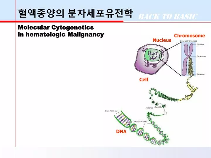
Chromosome. Nucleus. 혈액종양의 분자세포유전학 Molecular Cytogenetics in hematologic Malignancy. Cell. DNA. Historical Backgroud. Philadelphia chromosome (Ph) 1970 Banding technique 1970 t(9;22) in CML 1973 t(8;21) in AML-M2 t(8;14) in Burkitt lymphoma/Leukemia

E N D
Chromosome Nucleus 혈액종양의 분자세포유전학Molecular Cytogenetics in hematologic Malignancy Cell DNA
Historical Backgroud • Philadelphia chromosome (Ph) • 1970Banding technique • 1970 t(9;22)in CML • 1973 t(8;21)in AML-M2 • t(8;14)in Burkitt lymphoma/Leukemia • 1977 t(15;17)in AML-M3, t(4;11) • 1979 High-resolution banding technique • 1980 • 1982t(9;11) in AML-M5a, inv(3) in AML • 1983 inv(16) in AML-M4E • 1984 t(1;19) in ALL • t(1;3) in AML with dysmegakaryopoiesis • 1990 Molecular cytogenetics (FISH, CGH)
Cytogenetic & Gene Rearrangement • 1980 • 1983 MYC-IGH t(8;14) • 1984 BCR-ABL1 t(9;22) • IGH-BCL2 t(14;18) • 1990 • 1991 MLL-AFF1 t(4;11) • E2A-PBX1 t(1;19) • PML-RARA t(15;17) • RUNX1-RUNX1T1 t(8;21) • DEK-NUP214 t(6;9) • 1993 CBFB-MYH11 inv(16) • MLL-MLLT3 t(9;11) • 1997 TEL-AML1t(12;22)
KimSW, et al.: Banding patterns of normal human chromosomes. The Seoul Journal of Medicine 21(2):133-137(1980). Cytogenetic Studies for Hematologic Malignancies in Korea Table 4. Karyotypic pattern in cases with CML 조혈기질환에 있어서 골수세포 염색체 분석에 관한 연구. 대한혈액학회지 23(2),1988
Philadelphia Chromosome (=Ph) der(22)t(9;22)(q34.1;q11.21) q11.2 BCR q34 ABL Ph (formerly Ph1) may be used in text, but not in the description of the karyotype, where der(22)t(9;22)(q34;q11.2) is recommended.
50,XX,+8,t(9;22)(q34;q11.2),+10,+19,+der(22)t(9;22)(q34.1;q11.21)50,XX,+8,t(9;22)(q34;q11.2),+10,+19,+der(22)t(9;22)(q34.1;q11.21) CML Blast Crisis - Karyotype
Practical Use of Cytogenetics in CML • Advantages ofCytogentics, compared to molecular DNA studiesfor BCR gene rearrangement • Distinguish betwn variant Ph and standard t(9;22) • Dectect other abnormalities : +8, i(17q),+Ph,+19 • Predict or confirm blast crisis • Give information regarding percentage of normal vs abnormal cells • Valuable after BMT to follow engraftment of the donor cells and identify possible relapse
AML-M2 with t(8;21)(q22;q22) • Usually AML-M2, occasionally M1 & M4 • Young individuals with good remission rate • Blasts containing a single thin Auer rod • RT-PCR : RUNX1-RUNX1T1 fusion transcript
APL with t(15;17)(q22;q21), PML/RARA From highly fatal to highly curable RARA PML PML/RARA ATRA (all-trans retinoic acid) & ATO (arsenic trioxide) G-banding : t(15;17)(q22;q21) & Variants of t(15;Var;17), t(11;17), t(5;17) RT-PCR, FISH : PML/RARA fusion transcript
AML-M4Eo with inv(16) • Young patients, organomegaly, abnormal eosinophils • Specifically associated with M4Eo in over 50% of cases • Favorable prognosis, High incidence of CNS relapse MYH11 CBFB CBFB-MYH11
Acute Myeloid Leukemia and Related Neoplasms Proposed WHO Classification • AML with recurrent genetic abnormalities • t(8;21), RUNX1-RUNX1T1 • t(15;17), PML/RAR • inv(16), CBF/MYH11 • t(9;11), MLLT3/MLL • t(6;9), DEK/NUP214 • inv(3) or t(3;3), RPN1-EVI1 • t(1;22), RBM15-MKL1 • Acute myeloid leukemia with myelodysplasia-related changes • Therapy-related myeloid neoplasms • Acute myeloid leukemia, not otherwise specified • Myeloid sarcoma • Myeloid proliferations related to Down syndrome • Blastic plasmacytoid dendritic cell neoplasm
Normal Chromosome del 5q -7, del 7q del 9q del 20q +8 complex defects t(1;3), t(2;11) MDS t(3;3), inv(3) t(6;9), inv(16) t(8;21) t(9;22) t(v;11) t(15;17) AML
Myeloid Malignancy Secondary to Radiotherapy or Chemotherapy • Clinical features • AML-M1,M2,M6 • Lower remission rate and long-term reponse • Cytogenetics • Usually absence of t(8;21), t(15;17) & inv(16) • Usually complex karyotype • Common abnormalities: • -5, del(5q) in chemotherapy, • -7, del(7q) in radiation therapy, • Abnormalities of 3p, 11q23, 12p, and 17
Cytogenetics in Acute Lymphoblastic Leukemia • Poorly spread, short • Fuzzy chromosomes • Indistinct bands • Low success rate • 50% in conventional culture • 76% Clarkson (1985)
Ploidy Groups in Childhood ALL • Hyperdiploid >50 28% Early pre-B immunophenotype Found at the age of 2-10 years Lower WBC count, Favorable prognosis • Hyperdiploid 47-49 13% • Diploid 46 9% • Pseudodiploid 46 38% • Hypodiploid <46 6%
Precursor Lymphoid Neoplasm Proposed WHO Classification • B lymphoblastic leukemia/lymphoma, NOS • B lymphoblastic leukemia/lymphoma with recurrent genetic abnormalities • t(9;22)(q34;q11.2); BCR-ABL1 • t(v;11q23); MLL rearranged • t(12;21)(p13;q22) TEL-AML1 (ETV6-RUNX1) • hyperdiploidy • hypodiploidy • t(5;14)(q31;q32) IL3-IGH • t(1;19)(q23;p13.3);TCF3-PBX1 • T lymphoblastic leukemia/lymphoma
WHO Classification “Real” disease entity No single ‘gold standard’ Morphology Immunophenotype Genetic features Molecular & cytogenetics Clinical feature
AML with specific chromosome defects 1)HallymUniversityMedicalCenter (1995) 2)UniversityofMinnesotaMedicalSchool
Cytogenetics Technologies Karyotyping Provides a visual examination of the entire genome the best coverage but not the best resolution (≥ 5 Mb) Banding resolution differs from preparation to preparation FISH Sensitive (high resolution) Only provides information on tested regions, other aberrations will NOT be tested, i.e. not genome view M-FISH(Multi-FISH, Spectral karyotyping) Array-based Copy Number / CGH Analysis Single step global genome scan prevents FISHing expedition BAC (Bacterial Artificial Chromosome) Array Requires well characterized and high resolution clones High Resolution Oligonucleotide Microarray Highest resolution. Precise identification of gains and losses of genetic material.
Clinical Application of FISH • Microdeletion syndromes • Prader-Willi/Angelman syndrome • DiGeorge/CATCH 22 syndrome • Williams syndrome etc. • Marker chromosome • Hematologic malignancies • t(9;22): abl/bcr • t(15;17): PML/RARa • 11q23: MLL • t(12;21): TEL/AML1 • BMT follow-up using X/Y
Solid Tumor Cytogenetics 1. Preparation of tumor sample 2. Tumor cell culture Culture Media FBS (11-20%) Antibiotics Additives Tumor cells 2. Harvest 1) Colcemid (0.01-0.1mcg/mL) treatment 2) Hypotonic (0.050-0.075M KCl) treatment 3) Fixation 4) Slide preparation 3. Staining and analysis Insulin Transferrin Hydrocortisone EDGF
Cancer Cytogenetics (2009) • CONTENTS • 1) A New Approach to an Old Problem • 2) Cytogenetic Methods • 3) Cytogenetic Nomenclature • 4) Nonrandom Chromosome • Abnormalities in Cancer - An Overview • 5-10) Hematologic Malignancies • : AML, MDS, CML, CMPN, ALL, • Mature B- and T-Cell Neoplasms • and Hodgkin Lymphoma • 11-23) Solid Tumors : Upper Aerodigestive tract, • Lung, Digestive Tract, Urinary Tract, • Breast, Genital Organs, Endocrine Glands, • Nervous System, Eye, Skin, Bone, Soft Tissue Tumors