Download
1 / 1
10 likes | 224 Vues
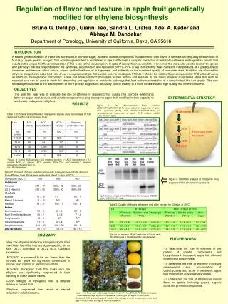
L-proline. glycine. malonyl-CoA. L-dimethylamino- phenylalanine. malonyl-CoA. malonyl-CoA. L-2-amino- butyric acid. malonyl-CoA. A-group (PII). B-group (PI). malonyl-CoA. L-threonine. L-pipecolic acid. malonyl-CoA. stimulates release of peptidyl-tRNA.

E N D
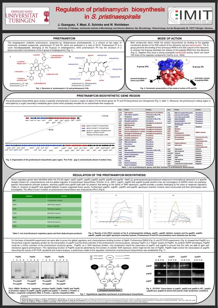
L-proline glycine malonyl-CoA L-dimethylamino- phenylalanine malonyl-CoA malonyl-CoA L-2-amino- butyric acid malonyl-CoA A-group (PII) B-group (PI) malonyl-CoA L-threonine L-pipecolic acid malonyl-CoA stimulates release of peptidyl-tRNA prevents binding of amino acyl-tRNA L-hydroxy- picolinic acid isobutyryl-CoA serine proline L-phenylglycine Literature: Bamas-Jacques, N., Lorenzon, S., Lacroix, P., de Swetschin, C. andCrouzet, J. 1999. Cluster organizationofthe genes ofStreptomycespristinaespiralisinvolved in pristinamycinbiosynthesisandresistanceelucidatedbypulsed-fieldgelelectrophoresis. Journal of Applied Microbiology 87: 939-948. Folcher, M., Gaillard, H., Nguyen, L.T., Nguyen, K.T., Lacroix, P., Bamas-Jacques, N., Rinkel, M., and Thompson, C.J. 2001.Pleiotropic functions of a Streptomycespristinaespiralisautoregulator receptor in development, antibiotic biosynthesis, and expression of a superoxide dismutase. J. Biol. Chem. 276: 44297-44306. Harms, J.M., Schlünzen, F., Fucini, P., Bartels, H. andYonath, A. 2004. Alterationsatthepeptidyltransferasecentreoftheribosomeinducedbythesynergisticactionofthestreptograminsdalfopristinandquinupristin. BMC Biology2:4. Regulation of pristinamycin biosynthesis in S. pristinaespiralis confirmed Positive regulation papR6 + PII PI suggested Positive regulation - + WT ∆papR1 ∆papR2 ∆papR4 24 48 72 96 24 48 72 96 24 48 72 96 24 48 72 96 papR3 - papR4 - papR5 C (mg/ml) C (mg/ml) papR4 J. Guezguez, Y. Mast, E. Schinkoand W. Wohlleben papR2 University of Tübingen, interfaculty Institute of Microbiology and Infection Medicine, Dpt. Microbiology / Biotechnology, Auf derMorgenstelle 28, 72076 Tübingen, Germany. + + + - papR1 Time (h) Time (h) + papR2 papR1 + + PRISTINAMYCIN MODE OF ACTION Pristinamycin Structural Genes Both compounds alone inhibit the protein biosynthesis by binding to the peptidyltransferase domain of the 50S subunit of the ribosome and are bacteriostatic.The A-grouppreventsthebindingoftheaminoacyl-tRNAtothe 50S subunitoftheribosome. In contrast, the B-groupfacilitatesthereleaseofthepeptidyl-tRNAfromtheribosome (Fig. 2). Together they show a strong synergistic bactericidal activity, which can reach 100 times of the separate components (Harms et al., 2004). The streptograminantibioticpristinamycin, produced by Streptomycespristinaespiralis, is a mixture of two types of chemically unrelated compounds: pristinamycin PI and PII, whichareproduced in a ratioof 30:70. Pristinamycin PI is a cyclic hexadepsipeptide, belonging to the B-group of streptogramins, while pristinamycin PII has the structure of a polyunsaturated macrolactone of the A-group of streptogramins (Fig. 1). I II 5‘ 3‘ 30S A P 50S pristinamycin IIA: dehydroproline pristinamycin IIB: proline pristinamycin IA: R = Me pristinamycin IB: R = H Fig. 2: Schematicpresentationofthemodeofactionof PI and PII. Fig. 1: Structure of pristinamycin I (I) and pristinamycin II (II). PRISTINAMYCIN BIOSYNTHETIC GENE REGION The pristinamycinbiosyntheticgeneclusterispartiallycharacterized. Itcovers a regionofabout 210 kbwhere genes for PI and PII biosynthesisareinterspersed (Fig. 3, table 1). Moreover, thepristinamycincodingregionisinterruptedby a crypticsecondarymetabolitegeneclusterwhichprobablyencodesfor an actinorhodin-likecompound. Fig. 3: Organizationofthepristinamycinbiosyntheticgeneregion. The 70 kb – gapisschematicallyshown in brokenlines. Table 1: List ofpristinamycin genes andtheirfunction in pristinamycinbiosynthesis. REGULATION OF THE PRISTINAMYCIN BIOSYNTHESIS Seven regulatory genes were identified within the 210 kb region: spbR, papR1, papR2, papR3, papR4, papR5 and papR6 . SpbR (S. pristinaespiralisbutyrolactone-responsive transcriptional repressor) is a specific receptor protein for γ-butyrolactones and the global regulator of pristinamycin biosynthesis (Folcheret al., 2001). papR1, papR2 and papR4encode proteins that are homologous to SARPs which are pathway-specific transcriptional activator proteins, whereas papR3 and papR5code both for proteins that belong to the family of TetR repressors. papR6encodes a protein belonging to the class of response regulators (Table 2). Analysis ofΔpapR1andΔpapR2deletionmutantssupportedtheseresults. FurthermorepapR3-, papR4-,papR5-andpapR6-apramycininsertionmutantswereconstructedandtheirphenotypeswereinvestigated. The effectofeachmutation on pristinamycinbiosynthesis was analyzedby HPLC (Fig.4). Fig. 4: Resultsofthe HPLC analysisoftheS. pristinaespiraliswildtype, papR1-, papR2- deletionmutantsandthepapR3-, papR4-, papR5-, papR6-andspbR- apramycininsertionmutants. Pristinamycin PI andPII concentrationswerefollowedoverthe time. Table 2: List ofpristinamycinregulatory genes andtheirdeducedgeneproducts. On thebasisofbandshiftexperimentswewereabletoprovethe global regulatoryandγ-butyrolactonebindingfunctionofSpbR. Furthermore EMSA (Fig. 5) andRT-PCR experiments(Fig. 6) showedthatPapR2 is a hierarchical superior regulatory protein for the transcription of papR1 and the direct activator of the pristinamycin structural genes, whereas PapR1 is a “helper” protein of PapR2. As another SARP homologue, PapR4 couldbe a furtheractivatorofthepristinamycinstructural genes . PapR5, as a TetRrepressorprotein, maytemporarilyretardtheexpressionofpapR1andpapR4 toensurethatthecellsareabletogainself-resistanceagainstpristinamycin. ThisrepressingfunctionofPapR5 couldbeabolishedbythefunctionofanotherTetRrepressor, whichmightbetheroleofPapR3. PapR6 mightcontrolthetranscriptionofpapR4.Assumingtheresultsof RT-PCR, bandshiftandmutantanalysis, a preliminary model oftheregulationmechanismofpristinamycinbiosynthesis was established (Fig. 7). PapR2 PapR4 PapR3 PapR5 1 2 3 4 1 2 3 4 1 2 3 4 1 2 3 4 Pro-papR1 Pro-papR1 Pro-papR4 Pro-papR1 Pro-papR2 Pro-papR3 Pro-papR5 Pro-papR4 Fig.5: EMSA: Binding ofregulatoryproteins PapR2, PapR4, PapR3 and PapR5 topromoterregionsofpapR1, papR3, papR4 andpapR5. 1: Control, 2: Protein, 3: Protein + unspecificcompetitive DNA, 4: Protein + specificcompetitive DNA. Fig. 6 : RT-PCR: TranscriptionofpapR1, papR2 andpapR4 in WT, ∆papR1, ∆papR2and ∆papR4 atseveral time pointsunderproductionconditions. Fig. 7 : Hypotheticalregulationmechanismofpristinamycinbiosynthesis.