Download
1 / 27
340 likes | 977 Vues
POSITRON EMISSION TOMOGRAPHY. Deepika Gupta 1 , Jayanti Tokas 2 , Shalini Jain 3 and Hariom Yadav 3* 1 Amity University, Noida, UP, India 2 Assistant Scientist, Biochemistry Department, COBS and Humanities, HAU, Hisar, Haryana , India 3 NIDDK, NIH, Bethesda, MD, USA

E N D
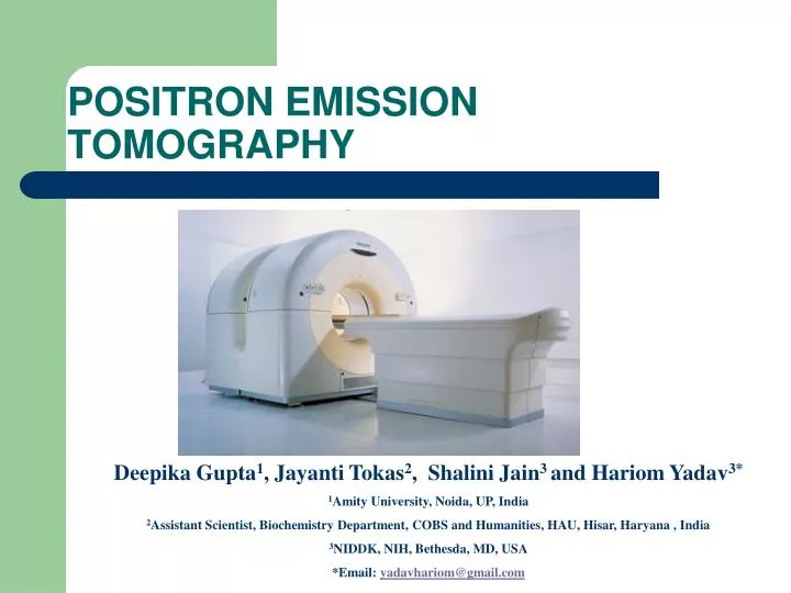
POSITRON EMISSION TOMOGRAPHY Deepika Gupta1, Jayanti Tokas2, Shalini Jain3 and Hariom Yadav3* 1Amity University, Noida, UP, India 2Assistant Scientist, Biochemistry Department, COBS and Humanities, HAU, Hisar, Haryana , India 3NIDDK, NIH, Bethesda, MD, USA *Email: yadavhariom@gmail.com
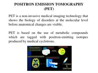
Definition A positron emission tomography is a nuclear medical imaging technique which produces a three dimensional image of functional processes in the body.
History of PET scan • The concept of emission and transmission tomography was introduced by David E. Kuhal and Roy Edwards in the late 1950s at the university of Pennsylvania. • In the 1970s, Tatsuo Ido at the Brookhaven National laboratory was the first to describe the synthesis of 18-F FDG, the most commonly used PET scanning isotope carrier. • Now there is not one person who developed the PET scan but a whole collection of people have made what it is today.
How it works A short lived radioactive tracer isotope, is injected in to the living subject (usually in to blood circulation) . The tracer is chemically incorporated in to a biologically active molecule. There is a waiting period while the active molecule becomes concentrated in tissues of interest. As the radioisotope undergoes positron emission decay (also known as positive beta decay), it emits a positron, an antiparticle of the electron with opposite charge.
After traveling up to a few millimeters the positron encounter an electron. The encounter annihilates them both, producing a pair of (gamma) photon moving in opposite directions. These are detected when they reach scintillator in the scanning device creating a burst of light which is detected by photomultiplier tubes. The technicians can then create an image of the parts of your brain, for example which are overactive.
Uses • Detect cancer. • Determine whether a cancer has spread in the body. • Assess the effectiveness of a treatment plan, such as cancer therapy. • Determine if a cancer has returned after treatment. • Determine blood flow to the heart muscle. • Determine the effects of a heart attack, or myocardial infarction, on areas of the heart. • Identify areas of the heart muscle that would benefit from a procedure such as angioplasty or coronary artery bypasssurgery (in combination with a myocardial perfusion scan). • Evaluate brain abnormalities, such as tumors, memory disorders and seizures and other central nervous system disorders. • To map normal human brain and heart function.
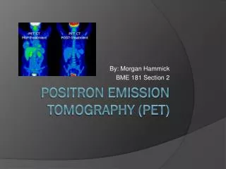
Combined PET/CT scanner • To detect structure and function simultaneously. • Greater detail with a higher level of accuracy; because both scans are performed at one time without the patient having to change positions, there is less room for error. • Greater convenience for the patient who undergoes two exams (CT & PET) at one sitting, rather than at two different times.
Tracer • Radioisotopes used in PET scans are isotopes of carbon, nitrogen,oxygen,gallium and 18F used as a substitute of hydrogen. • Only radioactive forms of natural elements that will pass safely through your body and be detected by the scanner. • The type of scanner used depends on what your doctor wants to measure. For example, if your doctor is looking at the tumor, he might use radio labeled glucose (FDG) and watch how it is metabolized by the tumor.
Cyclotron • Charged particle accelerator. • Accelerates charged particles in a cycle path and these particles gain energy. • Energetic particles then hit a target material get absorbed in to the nucleus, converting the target in to the different species. • For example, a proton of hydrogen, when hits 18O-water converts it to the 18F-fluoride with emission of a neutron other insignificant subatomic particles to balance the energy equilibrium.
Hormone • Chemical substances created by the body that control numerous body functions. • Biological compounds that communicate information at a distance. Hormones require specific receptors to begin their biological action use second messengers to initiate the cellular process that uses that information. • Substances secreted by various organs of the body that regulate growth, metabolism, and reproduction. They influence the growth and activity of cells. e.g Atrial natriuretic hormone, thyrotropin, growth hormone, androgens, insulin etc.
Receptors • Receptors bind with a substance (ligand) for which they are structurally shape specific. • Receptors can be found all over the place – inside a cell and specially embedded within and an integral part of all the membranes that a given cell may have. • Every function, response, interaction, pathway, and any other term you might think of that concerns the moment to moment existence of a cell, is controlled by various receptor/ligand-induced systems.
Neuroendocrine system • Endocrine system is a communication system in which hormones act as biochemical messengers. • Nervous system performs the same functions using electrical impulses as messengers. • Endocrine cells share a number of antigens with nerve elements, the term “neuroendocrine” is also used. neuroendocrine system is the combination of those two systems.
Neuroendocrine tumors • Heterogenous group of neoplasms • Originate from endocrine glands (pititary, parathyroids), • Neuroendocrine (adrenal), • Endocrine islets within glandular tissue (thyroid, pancreatic) • Cells dispersed between exocrine cells, such as endocrine cells of the digestive (GEP) and respiratory tracts. • Produce and secrete a variety of metabolically active substances (amines and peptides) • Cause distinct clinical syndromes, possess neuroamine uptake mechanisms and/or specific receptors at the cell membrane, such as somatostatin (SS) receptors, may occur either sporadically or as part of familial syndromes.
Peptide receptors expressed in GEP-NET • Somatostatin receptors • GLP-1 receptors • Secretin receptors • Cholecystokininreceptors • VIP receptors • Bombesin receptors • CRF receptors • CRF receptors • NPY receptors
Somatostatin • Somatostatin consists of a family of a 14-amino acid (somatostatin- 140 and a 28-amino acid (somatostatin-28) peptide. • Appears in sevsral organ systems such as central nervous system, the hypothalmopituitary system, the gastrointestinal tract,the excrine and endocrine pancreas and the immune system. • Somatostatin can be considered to be a neurotransmitter, a neurohormone or a local hormone acting via autocrine or paracrine mechanisms • Somatostatin and somatostatin analog inhibit tumor growth • Somatostatin has shorter half life (1-2 min) • Somatostatin analogs- octrotide and lanreotide has longer half life (1.5-2 h)
Newer analogues such as DOTA-Tyr3 octreotide (DOTATOC) have better uptake than octreotide. • The phenylalanine residue at position 3 is replaced by tyrosine, making the compound more hydrophilic and increasing the affinity for SSTR2, leading to higher uptake in SSTR2-positive tumours.
SS inhibits the proliferation of both normal and tumoral cells by • Hypophosphorylation of the retinoblastoma gene product • G1 cell cycle arrest • Apoptosis through SS receptor 3 induced by p53 and Bax • Inhibition of growth factors and angiogenesis
Somatostatin analog • DOTA-octreotide which has a very high affinity for SSTR2. • DOTA lanreotide has high affinity for SSTR5. • DOTA-1-NaI-octreotide(DOTANOC), which has shown a high affinity for SSTR2, SSTR3 and SSTR5. 3 somatostatin analogs, OC, TOC and TATE were conjugated to the metal chelator DOTA and labeled with the radiometal 111In, 90Y and 67Ga.
Somatostatin Receptors • Five human stomatostatin receptor subtypes (sst1, sst3, sst4, sst5) have been cloned and partially characterised. • Sst’s belong to the family of G protein coupled receptors characterised by seven transmembran domains. • Sst3 and sst2 internalize much better than sst1.
Octreotide • Brand name Sandostatin • An octapeptide that mimics natural somatostatin pharmacologically • Potent inhibitor of growth hormone, glucagon, and insulin than the natural hormone. • Since octreotide resembles somatostatin in physiological activities, it can: 1. Inhibit secretion of many hormones, such as gastrin, cholecystokinin, glucagon, growth hormone, insulin, secretin, pancreatic polypeptide, TSH, and vasoactive intestinal peptide. 2. Reduce secretion of fluids by the intestine and pancreas. 3. Reduce gastrointestinal motility and inhibits contraction of the gallbladder. 4. Inhibit the action of certain hormones from the anterior pituitary. 5. Cause vasocontriction in the blood vessels. 6. It has also been shown to produce analgesic effects, most probably acting as a partial agonist at the mu opiod receptor.
Chelating agent • An organic (hydrogen and carbon containing) compound that binds to charged metallic atoms (ions) to increase absorption. • A chemical compound that has the ability to bind strongly with metal ions. • Additive that can form several bonds to a metal ion, in order to deactivate them.Examples of chelating agents are: EDTA, ethylenediamine, phosphite. Syn. Complexing agent. DOTA ( 1,4,7,10-tetraazacyclododecane-N,N,,N,,,N,,,-tetraacetic acid) and DTPA ( Diethylenetriamine penta acetic acid) are two commonly used chelating agent in case of somatostatin.
Radiopharmaceuticals • Drugs containing a radioactive substance, used in the diagnosis and treatment of cancer and in pain management of bone metastases. • Medicinal products that are radioactive when used in patients. They are primarily used for diagnostic purposes. The radiation from the radiopharmaceuticals makes it possible to photograph the distribution of the medicinal product throughout the body. • It is the study and preparation of radiopharmaceuticals, which are radioactive pharmaceuticals.
Important radiopharmaceuticals for neuroendocrine tumor imaging (somatostatin receptors) Gallium + DOTA + NOC = 68Ga-DOTA-NOC =Radipharmaceutical ([1,4,7,10-tetraazacyclododecane-1,4,7,10-tetraacetic acid]-1-Nal3-Octreotide) 1,4,7,10-tetraazacyclododecane-N,N,,N,,,N,,,-tetraacetic acid (DOTA)0,Tyr3,octrotide (DOTATOC) DOTA0,Tyr3octreotate (DOTATATE) which has higher affinity for SSTR2.
Benefits of PET scan • The information provided by nuclear medicine examinations is unique and often unattainable using other imaging procedures. • For many diseases, nuclear medicine scans yield the most useful information needed to make a diagnosis or to determine appropriate treatment, if any. • Nuclear medicine is less expensive and may yield more precise information than exploratory surgery. • By identifying changes in the body at the cellular level, PET imaging may detect the early onset of disease before it is evident on other imaging tests such as CT or MRI.
Limitations of PET scan • Time-consuming. • The resolution of structures of the body with nuclear medicine may not be as clear as with other imaging techniques, such as CT or MRI. • PET scanning can give false results if chemical balances within the body are not normal. • Because the radioactive substance decays quickly and is effective for only a short period of time, it is important for the patient to be on time for the appointment and to receive the radioactive material at the scheduled time. • A person who is very obese may not fit into the opening of a conventional PET/CT unit.