Download
1 / 38
440 likes | 854 Vues
The Knee Joint. Anatomy and Physiology of Human Movement 420:050. Objectives. Bones, bony landmarks and joints Muscles Movements. Knee Joint. Large joint Complex ligamentous structures Femoral condyles articulate with tibial condyles

E N D
The Knee Joint Anatomy and Physiology of Human Movement 420:050
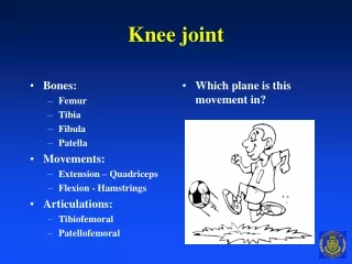
Objectives • Bones, bony landmarks and joints • Muscles • Movements
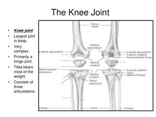
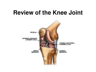
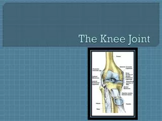
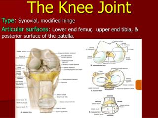
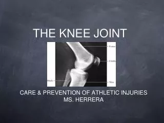
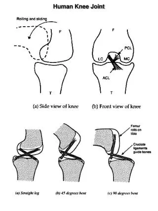
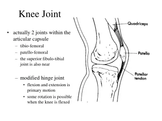
Knee Joint • Large joint • Complex ligamentous structures • Femoral condyles articulate with tibial condyles • Medial/lateral tibial condyles (aka plateaus) – act as receptacles for femoral condyles • Relatively stable joint despite injuries • Ligaments • Menisci • Quadriceps and hamstrings
Modified from Anthony CP, Kolthoff NJ: Textbook of anatomy and physiology, ed 9, St. Louis, 1975, Mosby.
Modified from Anthony CP, Kolthoff NJ: Textbook of anatomy and physiology, ed 9, St. Louis, 1975, Mosby.
Bones and Bony Landmarks • Tibia: • Bears weight • Fibula: • Serves as the attachment sight • Does not articulate with femur or patella • Not part of knee joint • Patella: • Sesamoid bone imbedded in patellar tendon • Improves mechanical advantage in knee extension • Bony landmarks same as the hip joint
Lateral femoral epicondyle Medial femoral epicondyle Patella Lateral tibial condyle Medial tibial condyle Head of fibula Tibial tuberosity
Joint • Knee joint • Diarthrodial uniaxial hinge joint • Movements • Planes and axes • Patellofemoral joint • Diarthrodial nonaxial gliding joint • Gliding nature of patella on femoral condyles
Objectives • Bones, bony landmarks and joints • Muscles • Movements
Quadriceps Rectus femoris Vastus medialis Vastus lateralis Vastus intermedius Hamstrings Biceps femoris Semimembranosus Semitendinosus Sartorius Gracilis Popliteus Gastrocnemius Muscles
Objectives • Bones, bony landmarks and joints • Muscles • Movements
Movements • Flexion • Bending or decreasing angle between femur and shin • Extension • Straightening or increasing angle between femur and shin
Movements • External rotation • Rotary movement of leg laterally away from midline • Internal rotation • Rotary movement of lower leg medially toward midline • Neither will occur unless flexed 20-30 degrees or more
FLEXION • Bending or decreasing angle between femur and shin
FLEXION • Biceps femoris • Semimebranosus • Semitendinosus • Sartorius • Gracilis • Popliteus • Gastrocnemius
EXTENSION • Straightening or increasing angle between femur and shin
EXTENSION • Rectus femoris • Vastus lateralis • Vastus medialis • Vastus intermedius