Download
1 / 1
10 likes | 103 Vues
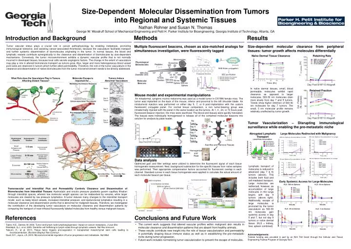
Size-Dependent Molecular Dissemination from Tumors in to Regional and Systemic T issues. 1 mm. *. Nathan Rohner and Susan N. Thomas George W. Woodruff School of Mechanical Engineering and Petit H. Parker Institute for Bioengineering, Georgia Institute of Technology, Atlanta, GA. *.

E N D
Size-Dependent Molecular Dissemination from Tumors into Regional and Systemic Tissues 1 mm * Nathan Rohner and Susan N. Thomas George W. Woodruff School of Mechanical Engineering and Petit H. Parker Institute for Bioengineering, Georgia Institute of Technology, Atlanta, GA * * * * Methods Results Introduction and Background * Multiple fluorescent beacons, chosen as size-matched analogs for simultaneous investigation, were fluorescently tagged Tumor vascular status plays a crucial role in cancer pathophysiology by enabling metastasis, promoting immunological tolerance, andassisting cancer-associated thrombosis, because the vasculature facilitates transport and further systemic dissemination of biomolecules originating in the tumor. In normal tissues, the blood and lymphatic vessels contribute synergistically to the clearance and dissemination of biomolecules by size-dependent mechanisms. Conversely, the tumor microenvironment exhibits a dynamic vascular profile that is not normally incurred in developed tissues, because local cells secrete angiogenic factors. The change in the extent of vasculature may play a role in altered biomolecule transport as tumors grow. Also, larger and more heterogeneous blood vessel pore sizes are observed in tumors which further alters permeability. Therefore, the role of the tumor vasculature in the clearance and dissemination of native biomolecules from the tumor microenvironment needs to be directly addressed. Size-dependent molecular clearance from peripheral tissues: tumor growth affects molecules differentially * * * * Naïve Dermal Tissue Clearance Retaining Rate Dextran Structure [Licor.com] NHS-Ester conjugation Polystyrene Structure SEC purification Molecular Escape is Important for… Tumors Induce Abnormal Vasculature What Role does the Vasculature Play in Tumors Affecting Distant Tissues? Mediating the Immune Response In naïve dermal tissues, small, blood permeable molecules exhibit rapid clearance as opposed to larger molecules. 500-30 nm molecules drain more slowly from day 7 and 9 tumors. Trends show higher retention of 500-30 nm molecules for day 7 tumors. The small, 5 nm molecular profile remains relatively unaffected by tumor growth. Naive Retained Fraction Mouse model and experimental manipulations An intradermal, syngenicmurine melanoma was used as a model tumor in C57/Bl6 female mice. The tumor was implanted on the back of the mouse, inferior and proximal to the left shoulder blade. An intratumoral injection was performed on either day 5, 7, or 9 post-implantation with the custom fluorescent conjugate panel. For normal tissue comparison, a non tumor-bearing group was intradermally injected with the panel in the same location as the tumors. At 1, 4 , 24, or 72 hours post-fluorescent beacon injection, the mice were sacrificed. The blood and tissues were quickly harvested. The tissues were individually homogenized to release all of the contained molecular beacons into solution for analysis by plate reader. Day 5 Tumor Randolph et al., Nat Rev Immunol.2005. Cancer-associated Thrombosis Day 7 Tumor Tatsumiet al., BBRC. 2013. Pre-metastatic Niche Formation Tobler and Detmar, J Leuk Biol. 2006. Day 9 Tumor No Tumor Tumor Vascularization – Disrupting immunological surveillance while enabling the pre-metastatic niche Quail and Joyce, Nature Medicine. 2013. Day 5 Tumor Day 7 Tumor Abrogated Lymphatic Transport Large Molecules Redirected with Malignancy Day 9 Tumor Sacrifice and Remove Tissues Homogenize and Plate PI + πI > PV+ πV PA > PI Data analysis Optimized gain and filter settings were utilized to determine the fluorescent signal of each tissue homogenate measurement. Next, background subtraction for the specific tissues from naïve samples was performed. Then appropriate compensation factors accounted for fluorescent overlap in each channel. Standard curves in each tissue homogenate were applied to calculate the actual amount of each molecular beacon per tissue. Lymphatic transport of molecules is reduced in advanced (day 7 & 9) tumors (above). This includes both fluid and cell-mediated transport. Large molecules are redirected, however, as accumulation of large molecules in the lungs begins with day 9 tumors (above, right). Additionally, escape of large molecules is promoted by the tumor vasculature as 500-30 nm molecules gain systemic access in day 5 and 7, but not day 9 tumors or naïve dermal tissue. (KLS = kidney, liver, spleen combined) 500 nm Sphere PI > PL Early Systemic Access for Large Molecules Interstitial Flux Transvascular Flux 5 nm Dextran (10 kDa) 50 nm Sphere 30 nm Dextran (500 kDa) Transvascular and Interstitial Flux and Permeability Controls Clearance and Dissemination of Biomolecules from Interstitial Tissues: Hydrostatic and oncotic pressure gradients govern capillary filtration through interstitial spaces, wherein low molecular weight species can be reabsorbed by venules, while larger molecules are cleared by low pressure lymphatics. A tumor induces many changes to the interstitial transport model, such as leaky blood vessels, increased interstitial pressure, and dysfunctional lymphatics resulting in a molecular clearance and dissemination profile that is abnormal for malignant tissues. Therefore, we investigated the contribution of the developing tumor vasculature on molecular clearance and dissemination patterns by comparing the transport of near-infrared fluorescently labeled molecules from healthy versus malignant tissues. -- Excitation ̶ Emission References Conclusions and Future Work • The current work suggests that altered vascular profiles within malignant skin results in molecular clearance and dissemination patterns that are absent from healthy animals. • These results contribute new insight into the role of tissue vascularization and permeability in potentially shaping tissue immune status as well as in establishing the pre-metastatic niche during tumor progression. • Future work includes normalizing tumor vascularization to prevent the escape of molecules. Tobler, N.E., Detmar, M. 2006. Tumor and lymph node lymphangiogenesis--impact on cancer metastasis. J Leuk Biol. Randolph, G.J., et al. 2005. Dendritic-cell trafficking to lymph nodes through lymphatic vessels. Nat Rev Immunol. Tatsumi, K., et al. 2013. Tissue factor triggers procoagulation in transplanted mesenchymal stem cells leading to thromboembolism. BiochemBiophys Res Commun. Quail, D.F., Joyce, J.A. 2013. Microenvironmental regulation of tumor progression and metastasis. Nat Med. Acknowledgements: Trainee funding was provided in part by an NIH T32 Grant through the Cellular and Tissue Engineering Trainee Program of Georgia Tech.