Download
1 / 22
220 likes | 799 Vues
THROMBOSIS. Dr. Afsar Saeed Shaikh M.B.B.S, M.Phil. Assistant Professor of Chemical Pathology Pathology Department, KEMU, Lahore. INTRODUCTION. NORMAL HEMOSTASIS 1) Maintain blood in fluid form in normal blood vessels

E N D
THROMBOSIS Dr. Afsar Saeed Shaikh M.B.B.S, M.Phil. Assistant Professor of Chemical Pathology Pathology Department, KEMU, Lahore.
INTRODUCTION • NORMAL HEMOSTASIS 1) Maintain blood in fluid form in normal blood vessels 2) induce a rapid & localized hemostatic plug formation at the site of vascular injury • THROMBOSIS ‘Pathologic opposite to hemostasis’
INTRODUCTION • DEFINATION: ‘An inappropriate activation of normal hemostatic processes, such as the formation of a blood clot in uninjured vasculature or thrombotic occlusion of a vessel after relatively minor injury.’
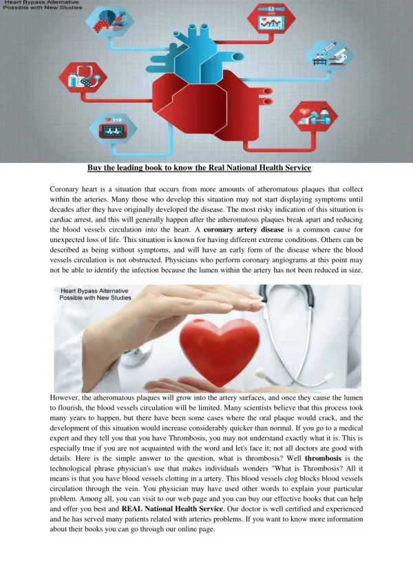
INTRODUCTION • ETIOLOGY: • Endothelial Injury • Abnormal Blood Flow • Hypercoagubality
1. Endothelial Injury • General: • A dominant influence • Can act without combination with other factors • Important factor where normally high flow rates hampers thrombus formation e.g. arterial circulation & heart chambers
Endothelial Injury • Sites : • Within cardiac chamber (e.g. following M.I) • Over ulcerative atherosclerotic plaques • At the site of inflammatory or traumatic vascular injury
Mechanism of Endothelial Injury • 1: Direct endothelial injury; physical loss of endothelium • 2: Dysfunctional endothelium (Imbalance of anticoagulant and pro-coagulant properties of endothelium) Continued…….
Dysfunctional Endothelium • Stress of hypertension • Bacterial endotoxins • Turbulent flow over scarred valves • Hypercholesterolemia • Products absorbed from cigarette smoke • Irradiation.
1. Abnormal Blood Flow • Turbulence: • Arterial & cardiac thrombosis • A cause of endothelial injury • Also causes countercurrents and local pockets of stasis • Stasis: • Venous thrombi • Acts by disturbing normal blood flow
Mechanism of Abnormal Blood Flow • Normal blood flow; laminar • Turbulence & stasis disrupt normal laminar blood flow • Bring platelets in contact with endothelium • Prevent dilution of clotting factors • Retard the inflow of inhibitors • Promote endothelial cell activation
Clinical Settings of Abnormal Blood Flow • Ulcerative atherosclerotic plaques • Aortic & arterial aneurysms • MI • Mitral valve stenosis • Hyperviscosity syndrome • Sickle cell anemia
3. Hypercoagubility • Important but less frequent contributor • ‘Any alteration of the coagulation pathways that predisposes to thrombosis’
Causes of Hypercoagubality • PRIMARY (Genetic) • Common: Mutation in factor V gene Mutation in prothrombin gene • Rare: Antithrombin III deficiency Protein C def. Protein S def.
Causes of Hypercoagubality • Secondary (Acquired) • High Risk: Prolonged bed rest MI, Cancer, DIC Atrial fibrillation Tissue damage Prosthetic cardiac valve Antiphospholipid antibody syndrome
Causes of Hypercoagubality • Secondary (Acquired) • Low Risk: Cardiomyopathy Nephrotic syndrome Pregnancy, Oral contraceptives Sickle cell anemia Smoking
Types of Thrombi • Types: • Arterial Thrombi • Venous Thrombi • Mural Thrombi • Red Thrombi (Stasis thrombi) • White Thrombi (Gray-white)
Morphology of Thrombi • Arterial: • Usually occlusive • Firmly attached to the injured artery wall • Gray-white and friable • Composed of a meshwork of platelets, fibrin, erythrocytes, and degenerating leukocytes
Morphology of Thrombi • Venous: • Invariably occlusive • Not firmly attached to the artery wall • Red in color and not friable but wet like a in-vitro clot • Contain more erythrocytes as compare to arterial thrombi
Fate of Thrombi • Propagation • Embolization • Dissolution • Organization and recanalization