Download
1 / 38
420 likes | 706 Vues
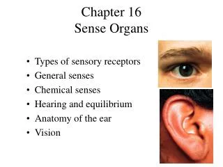
Chapter 15: Special Senses. Figure 15.23: Location and structure of taste buds on the tongue, p. 581. Taste fibers of cranial nerve. Gustatory hair. Epiglottis. Taste pore. Palatine tonsil. Lingual tonsil. Basal cell. Circumvallate papilla. Foliate papillae. Connective tissue.

E N D
Chapter 15: Special Senses
Figure 15.23: Location and structure of taste buds on the tongue, p. 581. Taste fibers of cranial nerve Gustatory hair Epiglottis Taste pore Palatine tonsil Lingual tonsil Basal cell Circumvallate papilla Foliate papillae Connective tissue Gustatory receptor cells Stratified squamous epithelium of tongue (c) Gustatory (taste) cells Taste pore Connective tissue Basal cells Taste bud (b) Fungiform papillae (a) (d)
Figure 15.24: The gustatory pathway, p. 583. Gustatory cortex (in insula) Thalamic nucleus (ventral posterior medial nucleus) Pons Solitary nucleus in medulla oblongata Facial nerve (VII) Vagus (nerve X) Glosso- pharyngeal nerve (IX)
Figure 5.21: Olfactory receptors, p. 579. Olfactory epithelium Frontal lobe of cerebrum Olfactory tract Mitral cell Olfactory tract Olfactory bulb Glomeruli Nasal conchae Cribriform plate of ethmoid bone Filaments of olfactory nerve Lamina propria connective tissue Route of inhaled air Olfactory gland Axon Basal cell Olfactory receptor cell Olfactory epithelium Supporting cell Dendrite Mucus Olfactory cilia Route of inhaled air containing odor molecules
Figure 5.22: Olfactory transduction process, p. 580. Extracellular fluid Na+ Odorant Adenylate cyclase Ca2+ 1 cAMP 2 3 GTP GTP 4 Receptor Golf GTP GDP 5 cAMP ATP Cytoplasm
George Wald 1906 – 1997 received the Nobel Prize in 1967 for discoveries concerning the primary physiological and chemical visual processes in the eye
Figure 15.1a: The eye and associated accessory structures, p. 557. Site where conjunctiva merges with cornea Eyebrow Eyelid Eyelashes Pupil Palpebral fissure Lacrimal caruncle Medial commissure (canthus) Lateral commissure (canthus) Sclera (covered by conjunctiva) Iris Eyelid (a)
Figure 15.1b: The eye and associated accessory structures, p. 557. Levator palpebrae superioris muscle Orbicularis oculi muscle Eyebrow Tarsal plate Palpebral conjunctiva Tarsal glands Cornea Palpebral fissure Eyelashes Bulbar conjunctiva Conjunctival sac Orbicularis oculi muscle (b)
Figure 15.2: The lacrimal apparatus, p. 558. Lacrimal gland Lacrimal sac Excretory ducts of lacrimal gland Lacrimal punctum Lacrimal canaliculus Nasolacrimal duct Inferior meatus of nasal cavity Nostril
Figure 15.3: Extrinsic eye muscles, p. 559. Trochlea Superior oblique muscle Superior oblique tendon Superior rectus muscle Axis at center of eye Lateral rectus muscle Inferior rectus muscle Conjunctiva Medial rectus muscle Lateral rectus muscle Optic nerve Inferior rectus muscle Inferior oblique muscle Annular ring (a) (b) Controlling cranial nerve Name Action Lateral rectus Moves eye laterally VI (abducens) Medial rectus Moves eye medially III (oculomotor) Superior rectus Elevates eye and turns it medially III (oculomotor) Inferior rectus Depresses eye and turns it medially III (oculomotor) Inferior oblique Elevates eye and turns it laterally III (oculomotor) Superior oblique Depresses eye and turns it laterally IV (trochlear) (c)
Figure 15.4a: Internal structure of the eye (sagittal section), p. 560. Ora serrata Sclera Ciliary body Choroid Ciliary zonule (suspensory ligament) Retina Macula lutea Cornea Fovea centralis Iris Pupil Posterior pole Anterior pole Optic nerve Anterior segment (cavity) Lens Scleral venous sinus (Canal of Schlemm) Central artery and vein of the retina Posterior segment (cavity) (contains vitreous humor) Optic disc (blind spot) (a)
Figure 15.6b: Microscopic anatomy of the retina, p. 562. Pigmented layer of retina Neural layer of retina Central artery and vein of retina Optic disc Sclera Optic nerve Choroid (b)
Figure 15.6a: Microscopic anatomy of the retina, p. 562. Pigmented layer of retina Horizontal cell Rod Cone Bipolar cells Amacrine cell Ganglion cells (a) Pathway of light
Figure 15.13: Focusing for distant and close vision, p. 567. Sympathetic + Nearly parallel rays from distant object Lens Ciliary zonule Inverted image Ciliary muscle Ciliary muscle Lens (a) Lens is flattened for distant vision Ciliary zonule (suspensory ligaments) Parasympathetic + Divergent rays from close object Inverted image (c) Anterior segment viewed from behind (b) Lens bulges for close vision
Figure 15.15: Photoreceptors of the retina, p. 570. Process of bipolar cell Light Light Light Synaptic terminals Inner fibers Rod cell body Rod cell body Nuclei Cone cell body Mitochondria Outer fiber Retinal (b) Opsin Inner segment Connecting cilia Apical microvillus Outer segment Discs being phagocytized Discs containing visual pigments Pigmented layer Pigment cell nucleus Melanin granules Basal lamina (border with choroid) (a)
Figure 5.19: Visual fields of the eyes and visual pathway to the brain, inferior view, p. 576. Fixation point Right eye Left eye Optic nerve Supra- chiasmatic nucleus Pretectal nucleus Optic chiasma Optic tract Lateral geniculate body Superior colliculus (sectioned) Uncrossed (ipsilateral) fiber Crossed (contralateral) fiber Lateral geniculate body of thalamus Optic radiation Superior colliculus Corpus callosum Occipital lobe (visual cortex) (a) (b)
Figure 15.25a: Structure of the ear, p. 584. Internal (inner) ear (labryinth) External (outer) ear Middle ear Auricle (pinna) Helix External acoustic meatus Lobule Pharyngotympanic (auditory) tube Tympanic membrane (a)
Figure 15.25b: Structure of the ear, p. 584. Entrance to mastoid antrum in the epitympanic recess Auditory ossicles Semicircular canals Malleus (hammer) Incus (anvil) Vestibule Stapes (stirrup) Vestibular nerve External acoustic meatus Cochlear nerve Cochlea Tympanic membrane Oval window (deep to stapes) Pharyngotympanic (auditory) tube Internal jugular vein Round window (b)
Figure 5.26: The three auditory ossicles in the right middle ear, p. 585. Malleus Incus Epitympanic recess Superior Anterior Pharyngotym- panic tube Tensor tympani muscle Tympanic membrane (medial view) Stapes Stapedius muscle
Figure 5.27: Membranous labyrinth of the internal ear, p. 586. Temporal bone Facial nerve Semicircular ducts in semicircular canals: Vestibular nerve • Anterior Superior vestibular ganglion • Posterior Inferior vestibular ganglion • Lateral Cochlear nerve Cristae ampullares in the ampullae Maculae Spiral organ (of Corti) Utricle in vestibule Cochlear duct in cochlea Saccule in vestibule Stapes in oval window Round window
Figure 5.29: Sound: source and propagation, p. 589. Area of compressed molecules Area of rarefaction Wavelength Crest Air pressure Trough (a) (b) Time Amplitude (c)
Figure 15.31: Route of sound waves through the ear, p. 590. External ear Middle ear Internal ear Air Malleus, incus, stapes (ossicles) External acoustic meatus Fluids in cochlear canals Oval window Tympanic membrane Pinna Lower Upper and middle Pressure Time Spiral organ (of Corti) stimulated One vibration Amplitude Amplification in middle ear
Figure 15.35: Structure of a macula, p. 594. Macula of saccule Macula of utricle Kinocilium Otolithic membrane Stereocilia Otoliths Hair bundle Hair cells Supporting cells Vestibular nerve fibers
Figure 15.36: The effect of gravitational pull on a macula receptor cell in the utricle, p. 595. Otolithic membrane Kinocilium Ster eocilia Depolarization Hyperpolarization Receptor potential (Hairs bent towar kinocilium) d (Hairs bent away from kinocilium) Nerve impulses generated in vestibular fiber Increased impulse frequency Decreased impulse frequency Excitation Inhibition
Figure 15.37: Location and sturcture of a crista ampullaris, p. 596. Flow of endolymph Crista ampullaris (a) Fibers of vestibular nerve Cupula (b) Turning motion Ampulla of right ear Ampulla of left ear Cupula Cupula at rest Position of cupula during turn Position of cupula during turn Fluid motion in ducts Horizontal ducts Increased firing Decreased firing (d) (c) Afferent fibers of vestibular nerve
Figure 15.38: Pathways of the balance and orientation system, p. 597. Vestibular receptors Visual receptors Somatic receptors Input Vestibular nuclear complex Reticular nuclei Cerebellum Central nervous system processing Oculomotor control (cranial nerve nuclei III, IV, VI) (eye movements) Spinal motor control (cranial nerve nuclei XI and vestibulospinal tracts) (neck movements) Output