Download
1 / 4
40 likes | 218 Vues
B. A. V. W. CS. Unmethylated. Methylated. I. E. 0. 0.25. 0.5. 1. 2.5. 5. 10. I. E. 0. 0.25. 0.5. 1. 2.5. 5. 10. A. C. 379. 134. 96. 72. 56. 43.

E N D
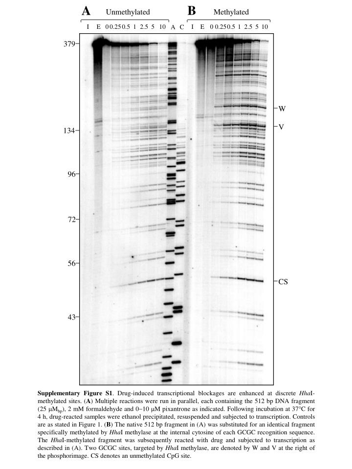
B A V W CS Unmethylated Methylated I E 0 0.25 0.5 1 2.5 5 10 I E 0 0.25 0.5 1 2.5 5 10 A C 379 134 96 72 56 43 Supplementary Figure S1. Drug-induced transcriptional blockages are enhanced at discrete HhaI-methylated sites. (A) Multiple reactions were run in parallel, each containing the 512 bp DNA fragment (25 Mbp), 2 mM formaldehyde and 010 M pixantrone as indicated. Following incubation at 37C for 4 h, drug-reacted samples were ethanol precipitated, resuspended and subjected to transcription. Controls are as stated in Figure 1. (B) The native 512 bp fragment in (A) was substituted for an identical fragment specifically methylated by HhaI methylase at the internal cytosine of each GCGC recognition sequence. The HhaI-methylated fragment was subsequently reacted with drug and subjected to transcription as described in (A). Two GCGC sites, targeted by HhaI methylase, are denoted by W and V at the right of the phosphorimage. CS denotes an unmethylated CpG site.
A B C Supplementary Figure S2. Quantitation of pixantrone-induced transcriptional blockages at sites V, W and CS of Supplementary Figure S1. The relationship between drug-DNA alkylation and pixantrone concentrations at two methylated CpG sites (V and W) and a single unmethylated CpG cite (CS) is presented in (A), (B) and (C), respectively. Unmethylated and methylated 512 bp DNA fragments are indicated by solid and open squares, respectively.
W V U CS 379 134 219 96 72 A B Unmethylated Methylated I E 0 0.25 0.5 0.75 1 1.25 A G I E 0 0.25 0.5 0.75 1 1.25 Supplementary Figure S3. Transcriptional blockages induced by formaldehyde-activated epirubicin are selectively enhanced at HhaI-methylated GCm5GC sites. (A) A native 512 bp fragment of DNA (25 µMbp) was initially reacted with 0–1.25 µM epirubicin (as shown) and 1 mM formaldehyde at 37°C for 4 h. Following an ethanol precipitation to remove unreacted drug, drug-reacted templates were subjected to transcription as detailed in the legend of Figure 1. Controls are as described in Figure 1. (B) The native 512 bp DNA fragment in (A) was substituted for an identical fragment specifically methylated by HhaI methylase at the internal cytosine of each GCGC sequence. This fragment was drug-reacted and processed as described in (A). U, V and W denote three GCGC sites. CS represents an unmethylated GpC site alkylated by formaldehyde-activated epirubicin.
A B D C Supplementary Figure S4. The frequency of epirubicin-adduct induced transcriptional blockages at GCGC sites U (A), V (B) and W (C) of Supplementary Figure S3. (D) The frequency of drug-induced blockages at site CS of Supplementary Figure S3, a GpC doublet not methylated by HhaI methylase. The mole fraction of transcript at each blockage site was determined for both unmethylated (solid squares) and HhaI-methylated (GCm5GC) (open squares) DNA templates.