Download
1 / 25
320 likes | 1.02k Vues
Hodgkin's Lymphoma. Introduction of lymphoma. The lymphomas are malignant tumors of lymphoid tissue ,characterized by the abnormal proliferation B or T cells in lymphoid tissue . Hodgkin’s lymphoma. Introduction:

E N D
Introduction of lymphoma The lymphomas are malignant tumors of lymphoid tissue ,characterized by the abnormal proliferation B or T cells in lymphoid tissue .
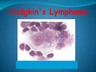
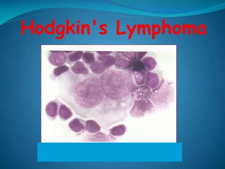
Hodgkin’s lymphoma Introduction: Hodgkin lymphoma (formerly called Hodgkin's disease) is a group of cancers characterized by Reed-Sternberg cells in an appropriate reactive cellular background. An important clinical feature is its tendency to arise within lymph node areas and to spread in an orderly fashion to contiguous areas of lymph nodes . Hodgkin lymphoma has a bimodal age distribution with one peak in the 20s and 30s, and a second peak over the age of 50.
Etiology Infection Genetics Occupational
Origin of Hodgkin's Lymphoma Evidence is now that the majority of classical HL have clonal Ig rearrangement, with somatic hypermutation clearly identifying that H-RS cells as a neoplastic, germinal center derived B- cells. Nodular lymphocyte predominant HL(NLPHL), as well is believed now to be derived from a B-cell as indicated by clonal VDJ rearrangements of the immunoglobulin heavy chain locus in the malignant lympocytic and histiostic cells (L & H-cells ).
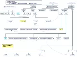
Histopathologic Classification WHO 1997 Nodular lymphocyte predominant Classical HL Lymphocyte rich CHL Nodular sclerosis Mixed cellularity Lymphocyte depletion Unclassified
Classical Hodgkin's lymphoma(CHL) • This variant comprises about 95% ,of all HL cases, and shows typical bimodal age distribution. • Each histological subtype is characterized by a series of clinical , morphological, phenotypic, and genetotypic features (nodular sclerosis, mixed cellularity ,lymphocyte depleted, and lymphocyte rich HL). • In CHL, classical RS-cells can be easily detected.
Diagnostic RS cells • Diagnostic RS-cells are large cells (20-50 Mm) in diameter or more with abundant weakly acidophilic or amphophilic cytoplasm, which may appear homogenous or granular and lacks a pale zone in the Golgi area , The nucleus is bilobed or polylobed . • The nuclear membrane is thick and sharply defined. The nuclear pattern is usually vesicular but with some coarse chromatin clumps scattered throughout .There is very large ,variously shaped, but usually rounded, highly acidophilic central inclusion like nucleolus surrounded by a clear halo. When the two lobes face each other (mirror image) owl eye appearance results. • Cells with this set of features but lacking nuclear lobation are referred to as mononuclear variant of RS cells or H-cells (Hodgkin's cells).
Nodular sclerosis Hodgkin's lymphoma (NSHL) Morphology In most cases, NSHL has at least a partially nodular pattern, with fibrosious bands separating the nodules. Areas of necrosis are common, and diagnostic RS- cells are rare. The characteristic neoplastic cells, the lacunar type of RS-cells, have multilobated nuclei and small nucleoli with abundant pale cytoplasm that retracts in formalin- fixed sections, producing an empty space or lacunae. The background usually contains several lymphocytes, histiocytes, plasma cells, eosinophils and neutrophils.
Mixed Cellularity Hodgkin's lymphoma(MCHL) Morphology The appearance is usually diffuse or vaguely nodular, without bands forming sclerosis. Fine interstitial fibrosis may be present, and classical diagnostic R-S-cells are easily identified. The background infiltrate typically consists of lymphocytes, epitheliod histiocytes, eosinophils, and plasma cells, but lymphocytes may predominate, giving rise to a differential diagnosis of nodular lymphocyte predominance Hodgkin's lymphoma(NLPHL) .
Lymphocyte depleted Hodgkin's lymphoma(LDHL) Morphology The pattern in LDHL is diffuse and often appears hypocellular ,due to the presence of fibrosis , necrosis, and a paucity of inflammatory cells. Large number of classical RS-cells are present with a bizarre pleomorphic sarcomatous variants. Confluent sheets of RS-cells may be present but rarely predominate .
Lymphocyte Rich Classical Hodgkin's Lymphoma (LRCHL) Morphology Some cases of the CHL with RS-cells of the classical type or lacunar type may have a background infiltrate that consist predominantly of lymphocytes, with rare or no eosinophils, the term lymphocyte rich CHL was proposed.
Nodular lymphocyte predominance Hodgkin's lymphoma(NLPHL) Morphology NLPHL is defined as having at least a partial nodular growth pattern, diffuse areas are present in a minority of the cases . The RS-cells variant of NLPHL differ from the "classic"RS-cells: they have vesicular, polylobated nuclei and distinct but small, usually peripheral nucleoli, without perinuclear halos, called L&H-cells (lymphocytes and/or histiocytes of Luke's and Butler) or "Popcorn cell". The classical RS are not usually found. The background is predominantly lymphocytes. Any clusters of epitheliod histiocytes, plasma cells, eosinophils and neutrophils are rarely seen.
Nodular LPHL and Classical HL Morphologic and Immunophenotypic Features.
General symptoms: • Fever • Weight loss • Loss of appetiteSymptoms of Hodgkin’s lymphoma: • Night sweats • Pruritus • lethargy
Examination • Local symptoms: • Enlarged, painless, non-tender, superficial lymph nodes • Alcohol-induced pain ــــRarely, patients with Hodgkin’s lymphoma complain of severe pain following alcohol ingestion. The pain typically occurs within a few minutes after the ingestion of even a small amount of alcohol. The mechanism is unknown.
Signs of HL: • Lymph node enlargement • Cachexia • Anemia • Splenomegaly • Hepatomegaly • Jundice. rarely
Diagnosis BLOOD: (FBC, Film, ESR, LFT, LDH, Urate , Ca.) Lymph node excision biopsy , image guided needle biopsy. Chest X-ray, CT of thorax, abdominal, pelvis and bone marrow biopsyـــــــstaging of HL