Download
1 / 26
290 likes | 538 Vues
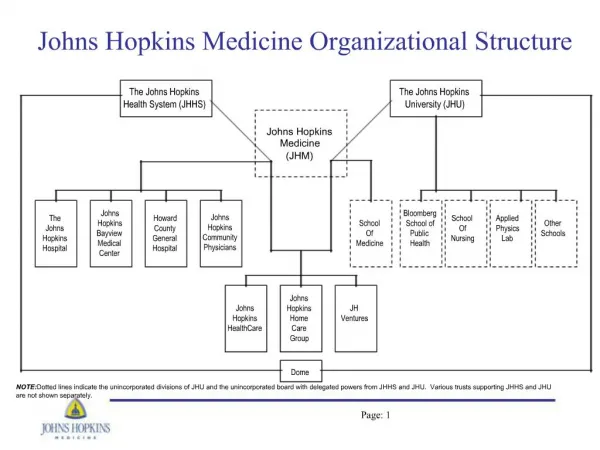
ETD & ETD/PTR. Electron Transfer Dissociation Proton Transfer Reaction. Page 1. y 3. y 2. z 2. y 1. z 1. z 3. b 1. c 1. b 2. c 2. b 3. c 3. ETD versus CID. ETD electron transfer surpasses internal heating rapid bond cleavage (no energy dissipation)

E N D
ETD & ETD/PTR Electron Transfer Dissociation Proton Transfer Reaction Page 1
y3 y2 z2 y1 z1 z3 b1 c1 b2 c2 b3 c3 ETD versus CID ETD • electron transfer surpasses internal heating • rapid bond cleavage (no energy dissipation) • random fragmentation of peptide backbone • leaves labile bonds like from PTMs intact • N-C bond cleavage yields c- and z-ion • preferable charge state z > 2 Conventional (resonant) CID • via several collisions with Helium precursor ion is internally heated • preferences for weak bond cleavages • nearby selected amino acids (E, D, P) backbone cleavage is preferred • b- and y-ions (and internal fragments) • best fragment spectra from 2+ ions Page 2
- n+ (n-1)+ ETD Reaction Scheme Multiply charged analyte(n≥ 2) Reagent radical anion odd-electron protonated peptide + Electron- transfer Cleavage of N-Cα bond Prerequisite: multiply charged precursor ions, n ≥ 2 ! ETD is not applicable to 1+ or negatively charged ions Page 3
ETD: No Cleavage at Proline Even though the N-C bond is cleaved no respective c and z fragments are formed since they stay connected via the Proline ring system. Page 4
The “3D Advantage” Non-linear Paul Trap: Dual injection and storage of ions of both polarities peptidecations & reagent anions Cations and anions are pushed towards the center of the trap Direct ETD reaction as soon as anions enter the trap Better cross sections for ion-ion-reactions in 3D trap due to compression into the same globular volume highly efficient ETD reaction Spec: ≥ 18 unique peptides from 5 fmol BSA on column (Easy-nLC) Page 5 Page 5
proteintermini Use of ETD for detailed Protein Characterization • Analysis of post-translational modifications (PTMs) • phosphorylation • glycosylation • deamidation etc. • Identification of sample preparation artifacts • MS/MS of large peptides • Combination of CID and ETD data for improved characterization of peptides and proteins, e.g. for QC applications. protein ID detailed characterization: PTM mixed modifications preparation artefacts Page 6 Page 6
No ! Yes ! Strategy for phosphopeptides: PTMScanTM PTMScanTM = neutral loss triggered ETD MS Loss of H3PO4:m = 98 Combination offast MS/MS for best sequence coverage (CID)anddetailed analysis of modified peptides (ETD) CID autoMS/MS Loss of Δm/z49, 32.6 and product ion among top N most intense MS/MS fragments ? ETD auto-MS2 of original intact PRECURSOR ion CID autoMS3 of neutral lossproduct ion(s) + Page 7 Page 7
Intens. x106 3+ 760.6 2+ 2.5 738.4 2+ 880.9 2.0 2+ 1.5 2+ 956.4 536.8 2+ 1.0 644.3 3+ 2+ 1+ 0.5 1+ 728.0 1141.0 445.1 1287.1 355.1 0.0 Intens. x104 3+ 722.0 loss of 32.6 6 3+ 728.0 4 triggers ETD MS/MSof 760.6 (3+) 1+ 2 1+ 1+ 1+ 440.3 1+ 844.4 932.4 1+ 303.2 1070.5 1166.7 0 m/z 200 400 600 800 1000 1200 1400 1600 PTMScanTM = Neutral Loss Triggered ETD phosphopeptide from asialo fetuin (tryptic digest) MS 3+ 760.6 Auto CID MS/MS Page 8 Page 8
Phosphoscan CID versus PTMScan ETD HTFSGVASVESSSGEAFHVGK, 2x phosphorylated, MW = 2279.9 Da from asialo fetuin (tryptic digest) CID: merged MS2 & pseudoMS3 CID MSn: Phosphorylation can not be assigned Page 9 Page 9
ETD ► Phosporylation at S11 and S13 Phosphoscan CID versus PTMScan ETD HTFSGVASVES*SS*GEAFHVGK, 2x phosphorylated, MW = 2279.9 Da from asialo fetuin (tryptic digest) ETD MS² Page 10 Page 10
Intens. 1+ 8 MS, 11.7 min x10 3+ 3+ 660.2 2+ 551.2 551.2 0.75 2+ 444.2 2+ 330.6 0.50 826.3 0.25 1134.7 1331.5 1495.2 7 x10 ∆m = - 35 → Neutral Loss of 105 Da CID (551.2) 3+ 4 516.3 CID : Almost no b- and y-ions ! 2 6 x10 3+ ETD (551.2) 2.0 ETD : Good fragment pattern ! 551.2 2+ 1.5 798.3 1.0 1+ 1+ 1+ 1+ 1+ 1+ 1+ 0.5 1146.4 918.3 1595.7 259.1 361.2 1020.5 1293.4 0.0 200 400 600 800 1000 1200 1400 1600 m/z Alternating CID-ETD for phosphopeptide analysis Identification of phosphorylation sites from a mixture of different caseins. ► Observation of several CID spectra showing a neutral loss of 105 Da instead of 98! Those spectra could not be identified via Mascot database search ? Page 11 Page 11
What causes a Neutral Loss of 105 Da ? A neutral loss of 105 Da can occur from carbamidomethylated methionine:1) carbamidomethylated methionine Loss of 105 Da Carbamidomethylation of methionine is a sample preparation artefact. It can be formed as side product during cysteine alkylation. 1) Krüger et al., Rapid Commun. Mass Spectrom. 2005; 19: 1709-1716. Page 12 Page 12
Mascot Database Search Results for α-S2-Casein Comparison of search results without and with modification Carbamidomethyl (M) without with modification Carbamidomethyl (M) ► With the knowledge of camMet as sample preparation artefact,two additional phosphopeptides are identified viaETD NcamMAINPpSKENLCSTCK & TVDcamMEpSTEVFTKK Page 13 Page 13
M* S* S* ETD Spectrum ofTVDcamMEpSTEVFTKK ETD of 551.2 (3+), tR = 11.8 min ► A single ETD spectrum allows for the identification of phosphorylation sites also in the presence of other labile modifications. Page 14 Page 14
Strategy for glycopeptide analysis • CID autoMS/MS analysis of the digested glycoprotein in enhanced resolution mode • Identification of the glycopeptides: • check for the presence of typical CID marker ions: - HexNAc: m/z 204 - HexNAcHex: m/z 366 - NeuAc: m/z 292, 274, 256 - HexHexNAcNeuAc: m/z 657 • only for O-glycans: check for neutral loss chromatograms, e.g. for hexose (54, 81, 162), HexNAc (101.5, 203), NeuAc (145.5, 291) • annotation of the sugar distances in order to determine the glycan residue • ETD experiment, either in autoMSn mode with or w/o inclusion list or in manual MS/MS mode to obtain best data quality. • Define the glycan moiety as modification in BioTools and match the ETD spectrum with the modified known sequence. Page 15 Page 15
pep N-acetylglucosamine galactose mannose fucose sialic acid pep 1360.7 pep pep 1229.0 pep pep 1200.2 pep pep pep 1098.9 pep 893.3 1506.7 pep 1709.8 1887.8 pep 2400.0 1025.6 944.9 * 366.1 528.2 1470.7 690.3 * 1046.5 1563.7 1157.6 * * 400 800 1200 1600 2000 2400 m/z Glycopeptide analysis using CID Fragments come almost exclusively from the cleavage of glycan moiety IgG3 tryptic digest glycopeptideMW 2602 Da Page 16 Page 16
[M+2H]2+ 1301.6 z9 z8 z7 z6 z5 z4 z3 2603.2 Side chain cleavage of N-glyc Asn Glu-Gln-Gln-Phe-Asn-Ser-Thr-Phe-Arg CH3 C=O NH z9. 2587.1 x 5 z4. z5. 2560.1 Asn z3. z8. z7. z6. 1099.4 408.2 2458.0 2330.0 708.4 2201.9 495.2 1041.4 687.5 927.4 2054.9 2403.1 516.3 m/z 400 800 1200 1600 2000 2400 Glycopeptide analysis using ETD Fragments arise from the cleavage of peptide backbone IgG3 tryptic digest Page 17 Page 17
ETD EQQFNSTFR CID Glycopeptide analysis using CID and ETD CID and ETD provide complementary information for glycopeptide identification Peptide Sequence Glycan moiety Page 18 Page 18
3+ c23 4+ c32 2+ c16 4+ c31 4+ (z+1) 31 c 1 (z+1) 1 m/z 800 805 810 815 3+ (z+1) 50 2+ (z+1) 33 Intens. 5 150 160 170 180 190 m/z x10 2.0 1696 1700 1704 m/z 1.5 2+ (z+1) 54 1.0 0.5 2780 2784 2788 m/z 0.0 500 1000 1500 2000 2500 m/z ETD analysis of large peptides galanin-like peptid (GALP)MWmono = 6200.3 Da multiply charged fragment ions up to z=4 are identified (Enhanced scan mode, 8100 m/z per sec) Page 19 Page 19
K G K T A G c H R G R G G W T L N S A G Y G P V L P S R G L G G z+1 R S H L L L Y S N L T W G G R H V P L A T K G K G G G E A z+1 59 z+1 23 c 38 c 23 z+1 37 z+1 22 c 60 z+1 32 z+1 31 z+1 33 z+1 29 c 33 c 20 z+1 28 z+1 53 c 31 c 32 z+1 19 z+1 27 z+1 52 c 30 c 19 c 27 z+1 50 z+1 36 z+1 26 z+1 30 z+1 51 z+1 48 z+1 15 z+1 34 c 26 c 22 c 50 z+1 58 z+1 47 c 13 c 6 z+1 41 c 8 c 16 z+1 25 z+1 57 z+1 21 z+1 46 c 12 c 28 c 35 c 37 c 40 c 15 c 48 c 4 z+1 43 z+1 24 z+1 56 c 21 c 45 c 11 c 36 c 34 c 39 c 7 c 14 c 43 c 9 z+1 44 c 10 c 3 c 57 z+1 38 c 25 z+1 54 c 5 z+1 42 c 17 z+1 49 3500 4000 4500 5000 5500 6000 500 1000 1500 2000 2500 3000 ETD of large peptides galanin-like peptid (GALP)MWmono = 6200.3 Da Deconvoluted spectrum m/z Page 20 Page 20
Use of ETD for detailed Protein Characterization protein ID detailed characterization: PTM • ETD-PTR top-down analysis for the determination of N- & C-termini ofintact proteins proteintermini mixed modifications preparation artefacts Page 21 Page 21
12+ MS 13+ 11+ 10+ Precusor Isolation [M+12H]12+ 12+ 9+ 500 1000 1500 2000 2500 m/z ETD 11+ 12+ 10+ m/z 500 1000 1500 2000 2500 9+ 500 1000 1500 2000 2500 m/z ETD & PTR for large peptides / small proteins Ubiquitin, bovine (8559.6 Da) Page 22 Page 22
ETD - PTR fragment charge states ≤ 6+ m/z 500 1000 1500 2000 2500 11+ PTRProton Transfer Reaction 12+ 10+ 9+ 500 1000 1500 2000 2500 m/z ETD & PTR for large peptides / small proteins Ubiquitin, bovine (MW = 8559.6 Da) Maximum Resolution Mode ETD fragment charge states ≤ 12+ Page 23 Page 23
+ Electron Transfer - 12+ n+ n+ n+ n+ PTR►Charge reduction using Proton Transfer Reaction + Proton Transfer - n+ m+ m+ m+ fragment ions with reduced charge states m = 6, 5, 4, 3, 2, 1 Principle of ETD-PTR Top Down Analysis ETD ► Production of highly charged fragment ions from intact proteins multiply charged fragment ions n =11, 10, 9, 8, ... Page 24 Page 24
- O O - • H - C PTR-reagents Benzoate anion (Hunt, Coon et al.) need two separate reagent reservoirs for ETD and PTR Perfluoro-1,3-dimethylcyclohexane = PDCH (McLuckey et al.) BrukerPTR reagent from fluoranthene + H C16H11- C16H10•- Page 25 Page 25
ETD & PTRfor large peptides / small proteins Ubiquitin, bovine (MW = 8559.6 Da) Deconvoluted spectrum Applications: e.g. QC of recombinant proteins, isolated proteins e.g. from cell lysates Advantages:no 1/3 cut-off, PTMs visible, good sequence coverage, N/C-termini included! Limitations:slow for LC separations, off-line techniques may be required (direct infusion, off-line nanospray,e.g. NanomateTM) Page 26 Page 26