Download
1 / 2
20 likes | 26 Vues
This hybridoma produces mAbs against Radial glia and neural progenitor cells.thttps://www.creative-diagnostics.com/Anti-Radial-glia-and-neural-progenitor-cells-hybridoma-137099-197.htm<br>

E N D
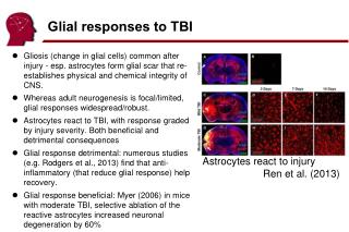
Mouse Anti-Radial glia and neural progenitor cells hybridoma [SD2] Anti-Radial glia and neural progenitor cells hybridoma Lot. No. (See product label) CELL LINE INFORMATION CSC-H1480 Radial glia and neural progenitor cells SD2 This cell line produces a monoclonal antibody RC1 (IgG class). Immunochistochemically, RC1 antibody reacts with radial glia and neural progenitor cells in CNS. Radial glial cells are a pivotal cell type in the developing central nervous system (CNS) involved in key developmental processes, from patterning and neuronal migration to their recently discovered role as precursors during neurogenesis. They arise early in development from neuroepithelial cells. Radial phenotype is typically transient, but some cells, such as Bergmann glia in the cerebellum and Muller glia in the retina, retain radial glia-like morphology postnatally. According to recent research, during the late stages of cortical development, radial glial cells divide asymmetrically in the ventricular zone to generate radial glial cells, postmitotic neurons and intermediate progenitor cells. Intermediate progenitor cells then divide symmetrically in the subventricular Neural progenitor cells are more promising candidates for replacing damaged and degenerated neurons because they are self- renewing, which allows for the in vitro production of many cells with minimal donor material. In order to confirm that the new neurons formed from neural progenitor cells are a part of a functional network, the presence of synapse formation is required. A study by Ma, Fitzgerald et al. is the first demonstration of murine neural stem and progenitor cell-derived functional synapse and neuronal network formation on a 3D collagen matrix. The neural progenitor cells expanded and spontaneously differentiated into excitable neurons and formed synapses; furthermore, they retained the ability to differenitate into the three neural tissue lineages. It was also demonstrated that not only active synaptic vesicle recycling occurred, but also that excitatory and inhibitory connections capable of generating action potentials spontaneously were formed. Thus, neural progenitor cells are a viable and relatively unlimited source for creating functional neurons. Radial glia and neural progenitor cells Mouse spleen Mouse x Mouse Hybridoma round Complete growth medium: RPMI1640 medium with 15% fetal bovine serum Atmosphere: air, 95%; carbon dioxide (CO2), 5% Temperature: 37.0 °C RPMI1640 medium with 15% fetal bovine serum Mycoplasma Status: Negative (MycoAlert Kit) Cat.No. Common Name Clone Cell Line Description Introduction Immunogen Immunological Donor Fusion Species Morphology Propagation Culture Medium Mycoplasma ANTIBODY INFORMATION Radial glia and neural progenitor cells N/A Target Application SAFETY AND PACKAGING liquid nitrogen Storage Creative Diagnostics. All rights reserved 45-16 Ramsey Road Shirley, NY 11967, USA Tel: 631-624-4882 ·Fax:631-614-7828 E-mail: info@creative-diagnostics.com www.creative-diagnostics.com
Safety Considerations The following safety precautions should be observed. 1. Use pipette aids to prevent ingestion and keep aerosols down to a minimum. 2. No eating, drinking or smoking while handling the hybridoma. 3. Wash hands after handling the hybridoma and before leaving the lab. 4. Decontaminate work surface with disinfectant or 70% ethanol before and after working with hybridoma. 5. All waste should be considered hazardous. 6. Dispose of all liquid waste after each experiment and treat with bleach. Dry Ice Ship Creative Diagnostics. All rights reserved 45-16 Ramsey Road Shirley, NY 11967, USA Tel: 631-624-4882 ·Fax:631-614-7828 E-mail: info@creative-diagnostics.com www.creative-diagnostics.com