Download
1 / 51
510 likes | 548 Vues
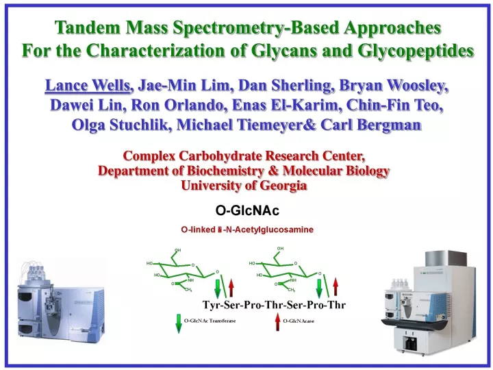
Tandem Mass Spectrometry-Based Approaches For the Characterization of Glycans and Glycopeptides. Lance Wells , Jae-Min Lim, Dan Sherling, Bryan Woosley, Dawei Lin, Ron Orlando, Enas El-Karim, Chin-Fin Teo, Olga Stuchlik, Michael Tiemeyer& Carl Bergman. Complex Carbohydrate Research Center,

E N D
Tandem Mass Spectrometry-Based Approaches For the Characterization of Glycans and Glycopeptides Lance Wells, Jae-Min Lim, Dan Sherling, Bryan Woosley, Dawei Lin, Ron Orlando, Enas El-Karim, Chin-Fin Teo, Olga Stuchlik, Michael Tiemeyer& Carl Bergman Complex Carbohydrate Research Center, Department of Biochemistry & Molecular Biology University of Georgia
Site Mapping and Characterization-Glycans, N-linked Sites, O-linked Sites P M Q N G S W E K A F S D Y P R S T P G L C N A I H T C I G R E S S Q L N S V K M Current Efforts: Glycopeptides
729.5 2 5 M a r 3 1 M # 2 1 8 3 R T : 3 5 . 9 4 A V : 1 N L : 1 . 5 6 E 7 T : + c N S I F u l l m s [ 3 5 0 . 0 0 - 1 7 5 0 . 0 0 ] 7 2 9 . 5 1 0 0 9 5 542.5 5 4 2 . 8 9 0 Peptide mixtures 8 5 HPLC 8 0 7 5 TurboSEQUEST Cross-Correlation Comparison Database 7 0 1082.7 6 5 1 0 8 2 . 7 6 0 Protein 5 5 Nucleotide ESTs 5 0 4 5 4 0 3 5 3 0 2 5 2 0 1486.1 1 5 1 4 8 6 . 1 1 0 Capillary Column Buffer A 5 Buffer B 0 4 0 0 5 0 0 6 0 0 7 0 0 8 0 0 9 0 0 1 0 0 0 1 1 0 0 1 2 0 0 1 3 0 0 1 4 0 0 1 5 0 0 1 6 0 0 1 7 0 0 LTQ ion trap Mass spectrometer Predicted MS/MS Nsi Peptides sequenced, Proteins Identified 1. Enolase EEALDLIVDAIK 2. P yruvate kinase NPTVEVELTTEK 3. Hexokinase IEDDPFVFLEDTDDIFQK 4. Hypusine APEGELGDSLQTAFDEGK 5. BMH1 QAFDDAIAELDTLSEESYK MS LC-nSI/MS/MS---Protein Identification Automated MS/MS CID MS/MS
The Glycan Problem??: Neutral Loss Fragmentation of GlcNAc-CTD by CID-MS/MS Parent ion 535 ([M+2H+] + GlcNAc) NL 7.62e6 y8 866.3 b ions - 164 251 348 449 536 633 720 848 100 Y--S--P--T--S--P--S--K 95 866 703 616 519 418 331 234 147 - y ions 90 Poor Fragmentation No Site Information 85 O-GlcNAc (204) 80 75 70 Relative Abundance 65 60 55 50 45 40 35 GlcNAc 30 y6 410.3 819.2 25 203.8 616.2 b2 20 b8 433.8 15 250.9 10 848.4 476.8 5 0 200 300 400 500 600 700 800 900 1000 1100 1200 m/z
Fragmentation of GlcNAc-CTD by CID-MS/MS/MS Parent ions 535/866 NL 2.88e6 b ions - 164 251 348 449 536 633 720 848 y6 b8 616.2 848.2 Y--S--P--T--S--P--S--K 50 866 703 616 519 418 331 234 147 - y ions Ion-Trap MS Allows MSn 45 Once sugar is off, fragmentation is good. MS/MS/MS works well for Sequencing, not site mapping 40 35 30 Relative Abundance 25 y3 331.0 20 830.2 15 598.2 y5 y4 b5 b7 10 518.0 804.3 y7 536.1 418.1 738.1 b6 720.1 499.9 y8 488.0 5 313.0 703.1 580.2 400.0 633.1 684.4 866.4 0 250 300 350 400 450 500 550 600 650 700 750 800 850 900 m/z
What We Really Need? • A method that allows for mapping of sites • A method that allows for enrichment of modified peptides • A method that can be used to do quantitative mass spectrometry (isotope labeling of PTM)
Michael Addition DTT (d0 or d6) H CH2 C N H C O Differential isotopic tagging of post-translationally modified ser/thr through b-elimination/Michael addition with light (d0) and heavy (d6) DTT. O CH2 C NH2 S b-Elimination H CH2 C N H C CH2 O C Alkylated Cysteine N H C Light DTT (d0) or Heavy DTT (d6) O OH OH Dehydroalanine (or (GlcNAc or phosphate) HSCd2CdCdCd2SH HSCH2CHCHCH2SH O OH OH b-Elimination H CH2 C N H C O O-GlcNAc or O-phosphate Modified Serine (or threonine)
LTQ: BEMAD (Light/Heavy) Quantified by LC-MS (200 amol) Theoretical: 1:1, Experimental: 1:0.88
Michael Addition DTT (d0 or d6) H CH2 C N H C O Differential isotopic tagging of both cysteine and post-translationally modified ser/thr through b-elimination/Michael addition with light (d0) and heavy (d6) DTT. O CH2 C NH2 S b-Elimination H CH2 C N H C CH2 O C Alkylated Cysteine N H C Light DTT (d0) or Heavy DTT (d6) O OH OH Dehydroalanine (or (GlcNAc or phosphate) HSCd2CdCdCd2SH HSCH2CHCHCH2SH O OH OH b-Elimination H CH2 C N H C O O-GlcNAc or O-phosphate Modified Serine (or threonine)
Comparison of Quantitative ICAT vs BEMAD * * ** *= site of phosphorylation, ** = site of O-GlcNAc Modification Quantification for both ICAT and BEMAD differed by <25% Collaboration with Keith Vosseller in Alma Burlingame’s Laboratory
BEMAD and Neutral Loss Approaches for Other O-Glycans • O-Man (not extended) on fungal proteins • Complex O-Glycosylation
N-linked glycosylation on Secreted Adipocyte ProteomeSite mapping using PNGaseF + 18O Water Sulfated glycoprotein 1 precursor (SGP-1) gi|3914939 (K)TVVTEAGNLLKDN#ATQEEILHYLEK(K)FSELIVNN#ATEELLVK (K)LVLYLEHNLEKN#STKEEILAALEK lipoprotein lipase gi|12832783 (R)TPEDTAEDTCHLIPGLADSVSNCHFN#HSSK vimentin gi|2078001 (R)QVQSLTCEVDALKGTN#ESLER Follistatin-related protein 1 precursor gi|2498391 (K)GSN#YSEILDK Haptoglobin gi|8850219 (K)VVLHPN#HSVVDIGLIK (K)NLFLN#HSETASAK (K)CVVHYEN#STVPEKK Adipsin gi|673431 (K)LSQN#ASLGPHVRPLPLQYEDK Decorin gi|6681143 (R)ISDTN#ITAIPQGLPTSLTEVHLDGNK Hemopexin gi|1881768 (R)SWSTVGN#CTAALR Cyclophilin C-associated protein MAMA/CyCAP gi|6755144 (K)GLN#LTEDTYKPR (R)ALGYEN#ATQALGR RIKEN cDNA 9330129D05 gene gi|30520293 (K)MELKN#QSRLQEPAAR Cathepsin L gi|929719 (R)AEFAVAN#DTGFVDIPQQEK PPBG/ Cathepsin A gi|12860234 (R)LDPPCTN#TTAPSNYLNNPYVR Relative Quantification Possible by 16O and 18O Water
Characterizing Plant Protein PGIP • N-linked glycoprotein • 7 Putative Sites of Glycosylation • Collaboration with Carl Bergmann’s and Mike Tiemeyer’s group at the CCRC
N-linked Site-Mapping on PGIP with PNGaseF Xcorr=4.82 gi|169684(K)IYGSIPVEFTQLNFQFLN@VSYNR@ = +3
PGIP N-linked Site Mapping with PNGase F/A PNGase F (red = coverage) 3 sites identified DLCNPDDKKV LLQIKKAFGD PYVLASWKSD TDCCDWYCVT CDSTTNRINS LTIFAGQVSG QIPALVGDLP YLETLEFHKQ PNLTGPIQPAIAKLKGLKSL RLSWTNLSGS VPDFLSQLKN LTFLDLSFNN LTGAIPSSLS ELPNLGALRL DRNKLTGHIP ISFGQFIGNV PDLYLSHNQLSGNIPTSFAQ MDFTSIDLSR NKLEGDASVI FGLNKTTQIV DLSRNLLEFN LSKVEFPTSL TSLDINHNKI YGSIPVEFTQ LNFQFLNVSYNRLCGQIPVG GKLQSFDEYS YFHNRCLCGA PLPSCK PNGase A (red=coverage) 7 sites identified DLCNPDDKKV LLQIKKAFGD PYVLASWKSD TDCCDWYCVT CDSTTNRINS LTIFAGQVSG QIPALVGDLP YLETLEFHK Q PNLTGPIQPAIAKLKGLKSL RLSWTNLSGS VPDFLSQLKN LTFLDLSFNN LTGAIPSSLS ELPNLGALRL DRNKLTGHIP ISFGQFIGNV PDLYLSHNQLSGNIPTSFAQ MDFTSIDLSR NKLEGDASVI FGLNKTTQIV DLSRNLLEFN LSKVEFPTSL TSLDINHNKI YGSIPVEFTQ LNFQFLNVSYNRLCGQIPVG GKLQSFDEYS YFHNRCLCGA PLPSCK High Stringency Filter
Glycan Release/Permethylation of Glycans • Trypsin digestion of a-DG • b-elimination- additon of NaOH • resulting in release of glycan structure from hydroxyl of Ser or Thr • Reduction with NaBH4 prevents re-attaching of glycan • Permethylation of glycan- OH→OMe • Addition of MeI • Analyzed permethylated glycans by applying MSn fragmentation as needed to completely determine the structure J. Am. Chem. Soc. (2003) 125(52): 16213-9.
PNGF PNGA LTQ Permethylated 1505.0 1331.7 MALDI
1331 PNGaseF M3N2X GlcNAc-OMe -Xyl, -H2O GlcNAc-OMe -GlcNAc- - MeOH
1505 PNGaseA
1054.55 M3N2XF PNGaseA GlcNAc-OMe - MeOH -Fuc or -Xyl-H2O -Fuc or -Xyl-H2O -GlcNAc- - MeOH
Site Mapping and Characterization-How unsatisfying for complex glycoproteins! P M Q N G S W E K A F S D Y P R S T P G L C N A I H T C I G R E S S Q L N S V K M
Alpha-Dystroglycana test case for mapping sites to structures
Fragmentation of O-glycans -SA -Hex -Hex -Hex-HexNAc + Na
MS3 – Pseudo Neutral Loss • Overall survey scan • 8 most intense peaks were selected • Fragmentation was induced upon these peaks • If result from fragmentation produced neutral loss, additional fragmentation was induced when the loss of a neutral from table was observed • Fragmentation was repeated resulting in MS3
MS Survey Scan MS survey scan MS survey scan MS survey scan MS/MS scan Neutral loss? No Yes Yes Top N peaks? No SEQUEST ID Yes MS/MS/MS scan Pseudo Neutral Loss Activated Data Dependant MS3 http://www.thermo.com
Use of Pseudo Neutral Loss Method Full Scan at 5.81 minutes
Neutral loss of SA MS/MS of 740.25
SA Hex HexNAC - SA - Hex (R)TPRPVPR(V) - HexNAc [M+2H]2+ [M+H]+ Identification of O-GalNAc Glycosylated Peptides
SA Gal GalNAC - SA - Gal (R)TPRPVPR(V) - GalNAc [M+2H]2+ [M+H]+ Assignment of Isobaric Glycans
Full Scan at 52.38 Full Scan at 52.38 minutes
MS/MS of 895.6 -SA
SA Hex HexNAc Hex (R)LETASPPTR(I) -Hex [M+H]+ [M+2H]2+ -HexNAc -Hex Identification of O-Man Glycosylated Peptides
SA Gal GalNAc Man (R)LETASPPTR(I) -Gal [M+H]+ [M+2H]2+ -GalNAc -Man Assignment of O-Man Isobaric Glycans
How can we isolate the exact site of glycosylation? • For peptide with only one Ser or Thr residue within sequence site of glycosylation is obvious • For peptides containing more that one Ser or Thr residue assignment is difficult… • Identify glycosylated peptide based upon addition of parent mass and combined neutral losses • Observe glycosylation of b & y ions • BEMAD-b-elimination and Michael addition of DTT
y6 y5 y3 y2 y1 (R)TP R P V P R (V) b2 b3 b4 b5 b6 [M+2H]2+ b2+HexNAc y4 b3+HexNAc [M+H]+ y4 y1 Identifying the Site O-GalNAc Glycosylation by b & y ions SA Hex HexNAC
Identifying the Site O-Man Glycosylation by b & y ions SA Hex HexNAc Hex -Hex (R)LETASPPTR(I) [M+H]+ b5+Hex b6+Hex [M+2H]2+ -HexNAc -Hex