Download
1 / 7
350 likes | 3.92k Vues
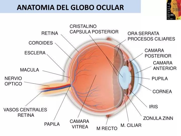
ANATOMIA DEL GLOBO OCULAR. CRISTALINO CAPSULA POSTERIOR. RETINA. ORA SERRATA PROCESOS CILIARES. COROIDES. CAMARA POSTERIOR. ESCLERA. CAMARA ANTERIOR. MACULA. NERVIO OPTICO. PUPILA. CORNEA. IRIS. VASOS CENTRALES RETINA. ZONULA ZINN. CAMARA VITREA. PAPILA. M. CILIAR.

E N D
ANATOMIA DEL GLOBO OCULAR CRISTALINO CAPSULA POSTERIOR RETINA ORA SERRATA PROCESOS CILIARES COROIDES CAMARA POSTERIOR ESCLERA CAMARA ANTERIOR MACULA NERVIO OPTICO PUPILA CORNEA IRIS VASOS CENTRALES RETINA ZONULA ZINN CAMARA VITREA PAPILA M. CILIAR M RECTO
VASCULARIZACIÓN ÓRBITA Y GLOBO OCULAR SENO CAVERNOSO V.OFTALMICA SUP V.CENTRAL RETINA V.OFTALMICA INF VENA LAGRIMAL A. LAGRIMAL VV. VORTICOSAS V.OFTALMICA SUP AA .CILIARES POST V. OFTALMICA INF SENO CAVERNOSO A. OFTALMICA A.CENTRAL RETINA V.CENTRAL RETINA LAT LAT
ANATOMÍA ECOGRÁFICA DEL GLOBO OCULAR Globo ocular, esférico. GO Cámara anterior, convexa, anecogénica. No valorable por eco. Cristalino, anecogénico salvo cápsula posterior. C Cámara vítrea, anecogénico. Contiene el humor vítreo. CV Pared posterior, contorno posterior ecogénico, liso y regular. PP Nervio óptico, cono hipoecogénico detrás del GO. NO Papila, prolongación anterior del NO en pared posterior. P Grasa retrobulbar, área ecogénica detrás del GO. Contiene el NO. GRB Glándula lacrimal, imagen hipoecogénicasuperolateral. GL Músculos extraoculares, haces hipoecogénicos detrás y lateral al GO. MM cv C GO Modo B PP PP Estudio Doppler color.
Disminuiremos la ganancia para valorar la órbita y su contenido. Es necesario aumentar la ganancia para valorar Adecuadamente la cámara vítrea. P MM NO GL GRB
VASCULARIZACIÓN ÓRBITA Y GLOBO OCULAR ARTERIAS ÓRBITA Y GO: Arteria oftálmica: Se adentra en la órbita a través de Foramen óptico, por fuera del NO. Tras pasar por encima del No se adentra antero medial en la órbita. AO. Sus principales ramas son: Vasos centrales de la retina: Arteria y vena central de la retina. Vascularizan los 2/3 internos de la retina. Se pueden localizar en el centro del segmento mas distal de NO. AVCR Arterias ciliares posteriores: Arterias ciliares posteriores largas y cortas. Vascularizan el 1/3 externo de la retina, coroides y la cabeza del NO. Penetran al GO medial y lateral al NO. ACP Arteria lacrimal. Para la glándula lacrimal. Arteria supraorbital. Arteria etmoidales, palpebral, frontal, angular, etc.. VIDEO AO
VASCULARIZACIÓN ÓRBITA Y GLOBO OCULAR ARTERIAS ÓRBITA Y GO: Arteria oftálmica: Se adentra en la órbita a través de Foramen óptico, por fuera del NO. Tras pasar por encima del No se adentra antero medial en la órbita. AO. Sus principales ramas son: Vasos centrales de la retina: Arteria y vena central de la retina. Vascularizan los 2/3 internos de la retina. Se pueden localizar en el centro del segmento mas distal de NO. AVCR Arterias ciliares posteriores: Arterias ciliares posteriores largas y cortas. Vascularizan el 1/3 externo de la retina, coroides y la cabeza del NO. Penetran al GO medial y lateral al NO. ACP Arteria lacrimal. Para la glándula lacrimal. Arteria supraorbital. Arteria etmoidales, palpebral, frontal, angular, etc.. VIDEO ACP AVCR
VASCULARIZACIÓN ÓRBITA Y GLOBO OCULAR VENAS ÓRBITA Y GO: Vena oftálmica superior, que drena al seno cavernoso. Se sitúa supero-medial en la órbita y es accesible al estudio Doppler en la mayoría de los pacientes. VOS Vena oftálmica inferior, que drena al plexo pterigoideo. Menos accesible. Ambos sistemas venosos se conectan por medio de colaterales. Venas vorticosas, se sitúan posterior al GO y drenan la coroides. VV VIDEO VOS VV VIDEO