Download
1 / 31
310 likes | 529 Vues
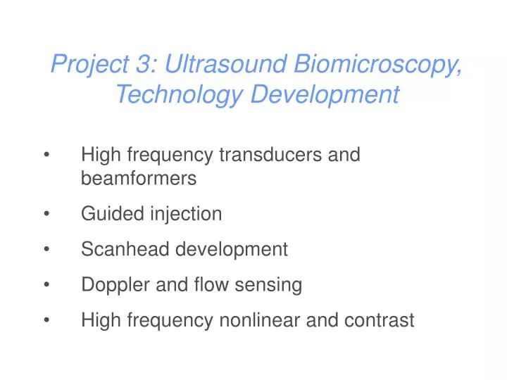
Project 3: Ultrasound Biomicroscopy, Technology Development. High frequency transducers and beamformers Guided injection Scanhead development Doppler and flow sensing High frequency nonlinear and contrast. Collaborating Groups. Commercial Partners.

E N D
Project 3: Ultrasound Biomicroscopy,Technology Development • High frequency transducers and beamformers • Guided injection • Scanhead development • Doppler and flow sensing • High frequency nonlinear and contrast
Collaborating Groups Commercial Partners MICe, Mt Sinai, Robarts, Queens, Univ of Virginia VisualSonics Commercial Collaborators AstraZeneca, Schering, Bracco, Imclone
Transducers and Beamformers Marc Lucaks, Jian Hua Yin, Richard Garcia, Guofeng Pang, Kasia Harasiewicz, Jeremy Brown Doppler / Flow Andrew Needles, Victor Yang Nonlinear / Contrast Manu Cherin, Ross Williams, Olivier Couture, Kevin Cheung
5 MHz 300 microns 30 microns 50 MHz
VisualSonicsVEVO 660 Mouse Scanner • B Mode (30 – 60) Hz • M Mode • Pulsed Doppler • EKV • 3D
Xenopus transverse section UBM - 30 fps 1 mm Yang, Needles et al U of Toronto
Micro-injection into the Midbrain 30 frames/s, RMV Probe
Chick embryo Day 3 Longitudinal Section Gharib/Brigande/Sahn/Thornberg labs: Cal Tech and Oregon Health and Sciences University
Chick embryo Day 3 Longitudinal Section Gharib/Brigande/Sahn/Thornberg labs: Cal Tech and Oregon Health and Sciences University
Array Development • Improve depth of field • Eliminate mechanical motion system • Improve system reliability
Annular Array Development Jeremy Brown, Geoff Lockwood
FPGA’s A/D’s
High Frequency Linear Array Development 5 MHz 300 microns 30 microns 50 MHz
Array Structure 75 microns SEM Images of Final Array Structure
Vendor : Gennum Material: Alumina ceramic Metal Layer : Au-8,000 Angs. Indium Solder: Indalloy 1E Dimension=20 x 4 x 2 mils Inner Electrodes: Bond pad Pitch= 150 um Bond pad opening = 70 um 1 mm Solder Reflow Richard Garcia and George Brown College
W:h = 0.43 35 Impedance of an Array Element
Doppler / Flow Imaging • Cardiogenesis in animal models • Cardiovascular Hemodynamics • Micro-circulation • Tumour blood flow
Imaging Tissue Motion: SAD map • Frame to frame clutter filtering with UBM at 15 fps R = Yang et al U of Toronto
Chick embryo Day 3 Longitudinal Section Yang et al U of Toronto
Ultrasound: Molecular Imaging Microbubble Contrast Agents Nanoparticle Contrast Agents • .5 – 5 microns • Gas filled • Inherently nonlinear • explodable • .1 – 1 microns • Liquid filled • Impedance enhanced signal
Microbubble Contrast Agents 6 Million frames/s Chin, deJong et al, Erasmus U Rotterdam 30 frames/s
3 min 15 min 15 min (E.M.) Albumin Microbubble Phagocytosis • Denatured albumin in microbubble shell adheres to activated leukocytes attached to inflamed endothelium • Albumin microbubbles remain acoustically active after phagocytosis Courtesy of Dr. JR Lindner, Circulation 2000;102
30 min Inflammation Targeted Microbubbles 2 h Post-ischemia inflammation seen with leukocyte targeted lipid microbubbles whose shell contains phosphatidylserine MB-PS Infarct (TTC) Christiansen JP, et al., Circulation 2002
Submicron Particles Intensity distribution of nanoparticles 500 nm • Liquid perflurocarbon, water, surfactant
1 mm Reflectivity dB) Distance in y Distance in x Ultrasound enhancement caused by the addition of submicron particles
Targeted contrast enhancement for ultrasound Contrast agent Probe
Project 3: Future Developments • Focus on array development • Special emphasis on beamformer • Improve sensitivity and quantitation for flow assessment • Develop, characterize and validate targeted contrast agents