Download
1 / 1
10 likes | 145 Vues
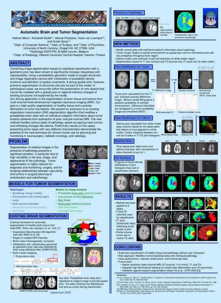
T1. Atlas. wm. wm. gm. Blue = outside red = traverse gold = inside cyan = exit purple = enter. gm. edema. tumor. csf. T2. distribution estimation. T1/T2 scatter plot. csf. T1 T2. Initialization. Step 3. Step 1. classification. Step 2.

E N D
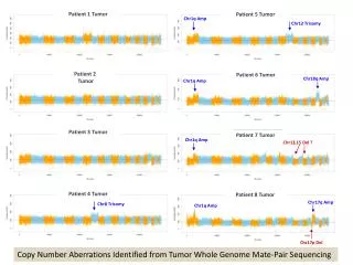
T1 Atlas wm wm gm Blue = outside red = traverse gold = inside cyan = exit purple = enter gm edema tumor csf T2 distribution estimation T1/T2 scatter plot csf T1 T2 Initialization Step 3 Step 1 classification Step 2 Presence of pathology Data: 3D MRI (1x1x1.5 and 1x1x3 mm3 voxels) T1 post T1 pre T2 EM segmentation using atlas of normals Automatic Brain and Tumor Segmentation Nathan Moon1, Elizabeth Bullitt2,1, Marcel Prastawa1, Koen van Leemput4,5, and Guido Gerig1,3 1 Dept. of Computer Science, 2 Dept. of Surgery, and 3 Dept. of Psychiatry, University of North Carolina, Chapel Hill, NC 27599, USA 4 Radiology-ESAT/PSI, B-3000 Leuven, Belgium 5 Dept. of Radiology, Helsinki University Central Hospital, Helsinki, Finland Classification fails in the presence of pathology NEW METHOD • Modify normal atlas with individual subject’s information about pathology • Tumor model: Region of partial enhancement as spatial prior and for estimating tumor pdf, low probability throughout brain region • Edema model: prior estimate of pdf and restriction to white matter region • Segmentation based on T1 pre-contrast and T2 channel only (T1 post only for atlas modif.) ABSTRACT Atlas Modification for Tumor p = 0.05 p = 1.0 Combining image segmentation based on statistical classification with a geometric prior has been shown to significantly increase robustness and reproducibility. Using a probabilistic geometric model of sought structures and image registration serves both initialization of probability density functions and definition of spatial constraints. A strong spatial prior, however, prevents segmentation of structures that are not part of the model. In pathological cases, we encounter either the presentation of new objects that cannot be modeled with a spatial prior or regional intensity changes of existing structures not explained by the model. Our driving application is the segmentation of brain tissue and tumors from multi-channel three-dimensional magnetic resonance imaging (MRI). Our goal is a high-quality segmentation of healthy tissue and a precise delineation of tumor boundaries. We present an extension to an existing expectation maximization (EM) segmentation algorithm that modifies a probabilistic brain atlas with an individual subject's information about tumor location obtained from subtraction of post- and pre-contrast MRI. The new method handles various types of pathology, space-occupying mass tumors and infiltrating changes like edema. Preliminary results on five cases presenting tumor types with very different characteristics demonstrate the potential of the new technique for clinical routine use for planning and monitoring in neurosurgery, radiation oncology, and radiology. - = T1 Difference Image Remap: Posterior Smoothness Constraints (MCF), base prob. Tumor prior calculated from the T1 pre- and post-contrast difference image. Mixture-model fitting gives a posterior probability of contrast enhancement. Difference intensities are remapped to a tumor probability image and smoothed (MCF). Posterior probab. tumor Multi-parameter fit Atlas Modification for Edema p = 0.1*wm Edema prior calculated from white matter atlas channel, based on the observation that edema is most apparent in white matter. Cluster initialized between wm and csf clusters in T1/T2 joint histogram. White Matter Atlas Edema Prior PROBLEM Modified Atlas wm gm csf tumor edema Final spatial prior adds tumor and edema channels, with normalization of atlas channels to sum to 1. Segmentation of medical images in the presence of pathology presents a significant problem, in particular due to high variability in the size, shape, and appearance of the pathology. Tumor segmentation is highly relevant for diagnosis and monitoring, surgery, and for studying relationships between vascularity and tumors in surgical planning of embolization and radiotherapy. EM Iterations Progress of tissue density distributions in joint histogram (top) and of classification (bottom). it 1 it 2 it 5 it 20 MODELS FOR TUMOR SEGMENTATION Radiologist • Something “wrong” is there • Enhancement with contrast agent • Lump • Multi-channel intensities • Deformation of normal anatomy • Asymmetry Models for Image Analysis • Probabilistic brain atlas, prior for normal • Use post-pre contrast difference • Blob Model • Multivariate statistical analysis • Atlas Deformation • Left-Right Deformation RESULTS T1 post T1 pre T2 3D view • Method has been applied to 5 cases • T1pre/T2 channels used for classification • T1 post (enhanced) helps initializing tumor cluster & prior • Partial volume errors due to coarse T2 resol. classification T1 post T1 pre T2 classification 3D view EXISTING BRAIN SEGMENTATION Existing framework for automatic segmentation of healthy brain tissue from head MRI: Koen van Leemput, et. al., KUL [1] T1 pre T2 classification 3D view T1 post bias estimation • Expectation Maximization EM algorithm (see also Wells et al. [2]) • Single or multiple MRI channels • Built in bias inhomogeneity correction • Initialization and classification governed by geometric prior (see also Warfield [3], here using statistical atlas (ICBM150 [4]) • Built in brain stripping CONCLUSIONS • Automatic classification of healthy tissue and pathology without user interaction • New approach: Modifies normal statistical atlas with individual pathology • Uses spatial priors, intensity model priors, and multivariate data • Future work: • Higher resolution triple channel MRI (3T scanner, 1x1x1mm3, T1 and T2) • Integrate high-dimensional warping to include atlas deformation and LR asymmetry • Validation against expert’s segmentation (Sean Ho et al., ICPR 2002 [5]) • Expectation step: • Maximization step: References: [1] van Leemput, K., Maes, F., Vandermeulen, D., Suetens, P.: Automated model-based tissue classification of MR images of the brain. IEEE TMI 18 (1999) 897-908. [2] W. M. Wells, W.E.L. Grimson, R. Kikinis, and F.A. Jolesz, Adaptive Segmentation of MRI Data, IEEE TMI 15(4):429-443, 1996. [3] S. Warfield, M. Kaus, F.A. Jolesz, and R. Kikinis, Adaptive template moderated spatially varying statistical classication,“ in MICCAI'98, Oct 1998, vol. 1496 LNCS, pp. 431-438, Springer. [4] ICBM Atlas, McConnell Brain Imaging Centre, Montréal Neurological Institute, McGill University, Montréal, Canada. [5] Sean Ho, Elizabeth Bullitt, Guido Gerig, Level Set Evolution with Region Competition: Automatic 3-D Segmentation of Brain Tumors, Proc. ICPR, IEEE, August 2002 Key idea: Probabilistic brain atlas [4] is registered to subject image to provide spatial prior. The atlas initializes the distributions and acts as a prior during classification ICBM150 MRI and tissue atlas September 2002