Download
1 / 16
160 likes | 369 Vues
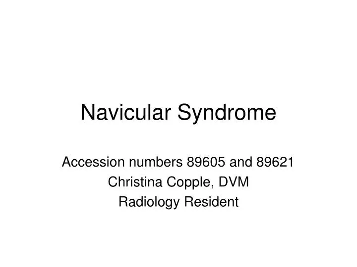
Navicular Syndrome. Accession numbers 89605 and 89621 Christina Copple, DVM Radiology Resident. Navicular Syndrome. Syndrome = group of symptoms, collectively indicate the presence of an underlying abnormal condition

E N D
Navicular Syndrome Accession numbers 89605 and 89621 Christina Copple, DVM Radiology Resident
Navicular Syndrome • Syndrome = group of symptoms, collectively indicate the presence of an underlying abnormal condition • Navicular Syndrome = chronically progressive, intermittent, bilateral forelimb lameness due to pain arising from the navicular apparatus • Can be unilateral and can occur in the hind limb
Navicular Apparatus • Navicular bone – distal sesamoid bone • Collateral ligaments • Distal sesamoidean impar ligament • Navicular bursa • Distal digital annular ligament • Deep digital flexor tendon - major compressive forces
4-navicular bone or distal sesamoid bone • 14- deep digital flexor tendon • 8- distal sesamoidean impar ligament • 16- navicular bursa • 15- distal digital annular ligament • 3- navicular bone or distal sesamoid bone • 8- deep digital flexor tendon • 6- distal sesamoidean impar ligament • 9a/9b- navicular bursa (proximal and distal recesses) • 11- distal digital annular ligament
Risk Factors • Concave or undulating proximal articular margin • Degenerative changes of palmar fibrocartilage • Physiologic vs. Non-physiologic • Fibrillation of DDFT • Enthesophytosis and mineralized fragments • Primary navicular bursitis http://www.portugalweb.com/images/Portuguese-horse-500-1.jpg
History, Clinical Signs and Diagnosis • Insidious onset, intermittent bilateral forelimb shifting leg lameness • Usually 7-9 yrs of age • Pain when standing at rest, pointing toe • Lameness more apparent moving in a circle with lame limb on inside • Usually positive response to palmar digital nerve blocks and navicular bursa analgesia • * No pathognomic findings • Diagnosis = characteristic gait, palmar foot pain, radiographic degenerative changes of navicular bone, rule-out other possible causes of lameness
Radiographs • Proximal border & Extremities • Enthesophytosis of extremities • Remodeling • Distal border • Synovial invaginations • Small osseous fragments • Flexor cortex • Cortical erosions • DDFT mineralization • Medullary Cavity • Radiolucent cysts • Sclerosis
Nuclear Scintigraphy • Patterns of increased radiopharmaceutical uptake commonly associated with palmar foot pain • Focal or generalized in navicular bone • Navicular bone and distal phalanx at region of insertion of DDFT • Navicular bone and palmar processes of distal phalanx • One or both palmar processes of distal phalanx • Distal phalanx at sites of insertion of DDFT
CT & MRI • Both more sensitive than radiographs to further characterize structural lesions of navicular bone, primary lesions of DDFT, identify degenerative changes of DIP articular cartilage • CT: osseous changes and surface contour of lateral wings, evaluate articular flexor surface • MRI: 3D spoiled gradient echo, 3D T2 gradient echo, STIR and fat-saturated 3D T2* • Soft tissues lesions – fibrocartilage changes, distension of navicular bursa, DDFT or impar ligament changes, abnormal bone marrow within navicular bone
SNORT • 16 yr old Quarter horse gelding • 3yr history of left forelimb lameness • Previously diagnosed with bilateral navicular “disease” • PE: right forelimb lame 3/5, positive on flexion tests in both forelimbs
Left Dorsolateral-Palmaromedial oblique Left Dorsomedial-Palmarolateral oblique
Left palmaroproximal-palmarodistal oblique Right palmaroproximal-palmarodistal oblique
Right dorsolateral-palmaromedial oblique Right dorsomedial-palmarolateral oblique
Right STIR Left STIR
References • Ross, Mike W., DVM and Sue J. Dyson, MA, VetMB, PhD, DEO, FRCVS. Diagnosis and Management of Lameness in the Horse. Saunders. Philadelphia, PA. 2003. • Thrall, Donald E., DVM, PhD. Textbook of Veterinary Diagnostic Radiology. 5th ed. Saunders. St. Louis, MO. 2007. • Stashak, Ted S., DVM, MS. Adams’ Lameness In Horses. 5th ed. Lippincott Williams & Wilkins. Philadelphia, PA. 2002. • Denoix, J.-M. The Equine Distal Limb Atlas of Clinical Anatomy and Comparative Imaging. Iowa State University Press. Ames, IA. 2000. • Pasquini, Chris, DVM, MS, Tom Spurgeon, PhD, and Susan Pasquini, DVM. Anatomy of Domestic Animals Systemic and regional approach. 7th ed. Sudz Publishing. Pilot Point, TX. 1996.