Download
1 / 69
720 likes | 1.13k Vues
Pharynx, Esophagus, Stomach Digital Laboratory. It’s best to view this in Slide Show mode, especially for the quizzes. This module will take approximately 60 minutes to complete. After completing this exercise, you should be able to:

E N D
Pharynx, Esophagus, Stomach Digital Laboratory It’s best to view this in Slide Show mode, especially for the quizzes. This module will take approximately 60 minutes to complete.
After completing this exercise, you should be able to: • Distinguish, at the light microscope level, each of the following: • Pharynx (oropharynx) • Esophagus • Upper • Middle • Lower • Stomach • Cardiac and pyloric regions • Pits and glands with mucus-secreting throughout (few chief or parietal cells) • Junctions • Esophageal-cardiac junction • Pyloric-duodenal (gastro-duodenal) junction • Pyloric sphincter • Body and fundus • Gastric pits • Surface mucous cells • Gastric glands • Mucous neck cells • Parietal cells • Chief cells • Distinguish, at the electron microscope level, each of the following: • Stomach • Chief cells • Zymogen granules • Parietal cells • Canaliculi • Tubulovesicles
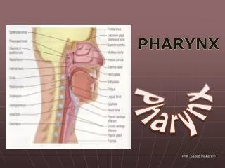
PHARYNX The pharynx is the crossing of the respiratory and digestive pathways. It has three parts, named for the structure that is anterior to that part: --nasopharynx is posterior to the nasal cavity --oropharynx is posterior to the oral cavity --laryngopharynx is posterior to the larynx The landmarks to officially demarcate these are the soft palate and epiglottis. This is just an overview FYI. You will look at these subdivisions and their distinctive characteristics when you study the respiratory system, and again in Brain, Mind, and Behavior, so don’t worry about the subdivisions now. What you want to focus on is the histology of the pharynx, and how that relates to swallowing. Our slide is from the oropharynx. lumen
PHARYNX Our slide of the pharynx is from the portion involved in swallowing. Although swallowing is a reflex action, the muscles within the pharynx are skeletal muscle. Many of these muscles have names (e.g. constrictor muscles) which you will learn in Brain, Mind, and Behavior. Being the conduit for boluses of food, the pharynx is lined by stratified squamous non-keratinized epithelium, and has substantial elastic tissue. lumen
PHARYNX The features of the pharynx seen in a low-magnification image include: Stratified squamous non-keratinized epithelium (black arrows) Lamina propria (black bracket), including of a dense band of elastic fibers (green bracket) Muscularis (purple bracket) consisting of skeletal muscle Note: no muscularis mucosa in the pharynx. lumen
PHARYNX Video of pharynx – SL96 • Link to SL 096 • Be able to identify: • pharynx
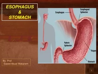
ESOPHAGUS The esophagus is a muscular tube that connects the pharynx with the stomach. It is the first portion of the digestive tract that has the four-layered structures we mentioned in the overview module: mucosa, submucosa, muscularisexterna, adventitia. As we mentioned before, the esophagus is in the posterior wall of the thorax, so the outer layer is an adventitia. lumen
ESOPHAGUS Characteristic features of the esophagus relate to it’s function: --epithelium is stratified squamous non-keratinized, providing a moist surface resistant to friction, conducive to movement of swallowed boluses toward the stomach --muscularisexterna transitions from skeletal (upper portion) to smooth muscle (middle and lower segments)….this is a gradual transition, so the upper-middle region contains a mixture of skeletal and smooth muscle --a thick muscularis mucosa --esophageal glands for lubrication
ESOPHAGUS Enlargement of the mucosa shows a stratified squamous non-keratinized epithelium and a thick muscularis mucosa (brackets). Esophageal glands are not readily apparent on this slide. Diffuse lymphoid tissue is not as prominent as in the rest of the GI tract; my guess is that this is because exposure to food is transient.
ESOPHAGUS Video of esophagus – SL16 Video of esophagus – SL15A • Link to SL 016and SL 015A • Be able to identify: • esophagus
ESOPHAGUS As mentioned already, the upper portion of the esophagus, being continuous with the pharynx, contains skeletal muscle in the muscularisexterna. This will transition into smooth muscle about 1/3 of the way toward the stomach, so that the muscularisexternain the lower portion of the esophagus is entirely smooth muscle. We do not have a section of the most cranial portion of the esophagus (horizontal line in drawing), but I hope you would be able to realize that an esophagus with a muscularisexterna composed of entirely skeletal muscle is from that region. The muscularismucosa has no such transition; it is always smooth muscle, from the cranial esophagus to the colon.
ESOPHAGUS Part way down the esophagus (about 1/3), smooth muscle begins to appear in the muscularisexterna. This creates a nice comparison of smooth and skeletal muscle that you looked at when first differentiating muscle types.
ESOPHAGUS By about the middle of the esophagus, the muscularisexterna has transitioned completely into smooth muscle. The remainder of the digestive tract will have smooth muscle in the muscularisexterna. Not a very attractive smooth muscle on this slide, but smooth muscle nonetheless.
ESOPHAGUS Video of middle esophagus – SL15A Video of middle esophagus – SL23 Video of lower esophagus – SL16 Don’t get confused here, the videos are not of “upper, middle, lower”. We don’t have an upper, so it’s two middles and a lower. • Link to SL 015A and SL 023 and SL 016 • Be able to identify: • Esophagus • Upper • Middle • Lower
STOMACH The stomach is a dilated portion of the GI tract that accepts food from a meal and slowly releases it into the duodenum. It also is involved in food breakdown, producing pepsin and HCl. Numerous mucous cells protect the mucosal lining from these harsh agents. As you are aware, the stomach can be divided into four major regions: --cardia --fundus --body --pylorus There’s also the pyloric antrum, pyloric part, etc., etc. Don’t get all in a tizzy about these for histology….
STOMACH HISTOLOGICALLY, we can divide the stomach into three parts: --cardia --fundus (which includes fundus and body) --pylorus These are demarcated by the dotted lines in the image. The cardia and the pylorus region are similar to each other, as are the body and fundus. We will discuss the cardia and pylorus first, along with their adjacent organs you already learned about, the esophagus and duodenum, respectively. After this, we will consider in detail the histological features of the fundus and body. Fundus here refers to the histological fundus, which includes the body. Before we do all that, lets overview some general features of the stomach…..
STOMACH Like the esophagus and intestines, the stomach has the four-layered structure characteristic of the gastrointestinal tract. Features unique to the stomach include: --Rugae are internal folds in an empty stomach, which are not present when distended. These are large and not readily apparent on our slides. --The muscularisexterna consists of three layers of smooth muscle, an inner circular layer, outer longitudinal layer, and an oblique layer. This organization is not obvious on our slides. --The mucosa has a unique structure described on the next slide…..
STOMACH The surface of the stomach is relatively smooth (i.e. it lacks villi). There are openings of the internal surface that lead to deep holes called gastric pits. The inferior portion of each pit is a narrowed isthmus. Projecting from the bottom of the pits are two or more gastric glands. The gland can be divided into a neck and fundus (base), terms which will be used to help identify predominant cell types in those regions. The glands are tightly packed, with little lamina propria between them; as we will see, this often distorts your perception of these glands. The micrograph on the left is a nice longitudinal section through the pits and gastric glands. In this image, a pit/gland unit is outlined, with arrows showing continuity between the single pit and two of its glands.
STOMACH Here are three images from our slide set. All are cut in longitudinal section. The approximate extent of the pits (black brackets) and glands (blue brackets) is indicated in each image. The pits are fewer and wider than glands; this can be used to your advantage to determine the transition point, which is where the structures become narrower and more numerous. Note the depth of the pits varies in different regions of the stomach.
STOMACH These images show oblique sections through the mucosa (muscularis mucosa indicated by the arrows in the lower image). Even though the longitudinal views shown on the previous slide make is easier to see the pits and glands, you can certainly use the diameter of the lumen to determine the transition from pits to glands (dotted lines).
STOMACH Video of an overview of the stomach – SL10 Video of an overview of the stomach – SL48A Video of an overview of the stomach – SL73 • Link to SL 010 and SL 048A and SL 073 • Be able to identify: • Stomach • Pits • Glands
STOMACH – CARDIA AND PYLORUS As we mentioned, the cardia and pylorus regions of the stomach are quite similar. Apart from stem cells and enteroendocrine cells (which we don’t see on routine stain anyways), the epithelium of the pits and glands consists of mucus-secreting cells. cardia pylorus These mucus-secreting cells in the stomach (and in the gall bladder and pancreatic ducts) are more eosinophilic than what you have seen in the salivary glands and goblet cells; they are mucus-secreting cells nonetheless.
STOMACH – CARDIA AND PYLORUS Subtle histology-geek note: You can tell the difference between the pyloris and cardia of the stomach by comparing the height of the pits and glands (i.e. pit-to-gland ratio). cardia pylorus This difference is easy to see if you have them side-by-side. However, this might not always be the case. Fortunately, I have a tendency to use these regions on exams with their adjacent organs, so you will see “esophageal-cardiac junction” and “pylorus-duodenal junction”.
ESOPHAGEAL-CARDIAC JUNCTION Video of the esophageal-cardiac junction – SL48A Video of the esophageal-cardiac junction – SL48B • Link to SL 048Aand SL 048B • Be able to identify: • Esophageal-cardiac junction
PYLORUS-DUODENUM JUNCTION Video of the pylorus-duodenum junction – SL73 • Link to SL 073 • Be able to identify: • Pylorus-duodenum junction
STOMACH – FUNDUS AND BODY • The mucosa of the histological fundus (fundus and body) of the stomach is the site of digestion. As with the rest of the tract, this region has stem cells and enteroendocrine cells. What is unique about this region: • The mucus-secreting cells cover the surface and line the pits (surface mucous cells), and some extend into the neck region of the glands (mucous neck cells) • Two new cell types are within the glands: • Parietal cells secrete HCl and gastric intrinsic factor. HCl activates pepsinogen, and is bacteriostatic. Intrinsic factor significantly increases the absorption of vitamin B12, which is necessary for RBC production. Parietal cells are more numerous in the upper portion of the glands. • Chief cells secrete pepsinogen, an inactive protease that is converted to an active form (pepsin) by HCl in the stomach lumen. Chief cells are more numerous in the base of the glands. So, basically what we’re saying here is, when compared to the cardia and pyloris, which have mucus-secreting cells throughout the pits and glands, the fundus and body have two additional cells that can be found in the glands, so there are not as many mucus-secreting cells in those glands.
STOMACH – CHIEF CELLS Lets look at drawings and EMs of chief and parietal cells to understand their function first; this will help explain their histological features on H&E-stained sections. Chief cells are pretty straightforward because they are protein-secreting cells. Therefore, you would expect to see plenty of rough endoplasmic reticulum, a prominent Golgi apparatus (or at least a place for one), and secretory granules, which are called zymogen granules. Hmmmm….. So, I’m thinking if I see an EM from a gland of the stomach, and a cell has these organelles in large quantities, then it’s a chief cell. I’m also thinking that in H&E sections, the RER will give the chief cell cytoplasmic basophilia on its basal aspect, and the secretory granules will stain eosinophilic at the apical end of the cell.
STOMACH – CHIEF CELLS Secretory granules In this EM from a gastric gland, the lumen is indicated by the “L”. At least three chief cells are shown across the top and right, the nucleus of one is indicated by the C. Part of a chief cell from an adjacent gland is in the lower-left. Note the elaborate rough endoplasmic reticulum in the basal aspect of these cells, and secretory granules (aka zymogen granules) clustered near the lumen. Secretory granules L Ignore me for now Secretory granules We’ll hold off looking at the light microscope for now until we describe the parietal cells. C
STOMACH – PARIETAL CELLS Secretion of HCl by parietal cells is done via membrane-bound transport channels. These channels are placed into an elaborate invagination of the plasma membrane called an intracellular canaliculus, which have numerous microvilli. These cells also have a tubulovesicular system that provides a reservoir of membranes with channels (see next slide). The channels are made “ahead of time” (see next slide), so the cell does not display elaborate rough endoplasmic reticulum. There are numerous mitochondria to support the energy requirements of the pumps, so these cells are eosinophilic on H&E.
STOMACH – PARIETAL CELLS This drawing is a of a single cell, split to show the cell in its inactive state (lower left) and active state (upper right). As mentioned, the cell produces the proton and chloride pumps “in advance”, and sequesters them in tubulovesicles near the apical membrane of the cell when the cell is inactive. Therefore, when the cell is inactive, it contains many of these tubulovesicles, and its intracellular canaliculus and microvilli are not well developed. As food enters the stomach, requiring acid secretion, the tubulovesicles fuse with the apical plasma membrane, allowing the membrane-bound channels to pump H+ and Cl- into the lumen of the stomach. This greatly enhances the depth of the intracellular canaliculus and the number of associated microvilli (while at the same time reducing the number of tubulovesicles). Hmmmm….. So, I’m thinking if I see an EM from a gland of the stomach, and a cell has lots of microvilli, a canaliculus, and lots of mitochondria and tubulovesicles, it’s a parietal cell. I’m also thinking that in H&E sections, the numerous mitochondria and tubulovesicles will impart cytoplasmic eosinophilia onto the parietal cell, and the cell will be large and round, with a centrally-located nucleus.
STOMACH – PARIETAL CELLS In this EM from a gastric gland, the lumen is indicated by the “L”. The nucleus of a parietal cell is indicated (PC). Note the two profiles of the intracellular canaliculus (IC) with their elaborate microvilli. This cell is filled with numerous tubulovesicles (a region with about 30-50 tubulovesicles is outlined in red). The dark oval structures in this cell are mitochondria. IC L PC It’s low power, but you can still compare the mitochondria to the secretory vesicles. The secretory vesicles are rounder, not as dark (though this varies), are clustered near the lumen, and within a cell with lots of rough endoplasmic reticulum. IC
STOMACH – FUNDUS AND BODY To look at these cells on glass slides, it’s easier to start with PAS-stained slides counter stained with eosin and azure (azure shows basophilia). In these images, you can see that: mucus-secreting cells (black outlines) are PAS positive parietal cells (green outlines) are large, with a central nucleus and eosinophilic cytoplasm chief cells (yellow outlines) have basophilia in their basal aspects and eosinophilia in their apical aspects Single cells are outlined. Outlines are animated so you can toggle back and forth. Parietal cells are eosinophilic and are more numerous in the upper gland, while chief cells are basophilic and are mostly in the base of the glands. Therefore, at low power (left) the top of the gland is more eosinophilic, the base is more basophilic.
STOMACH – FUNDUS AND BODY Video of the fundus and body PAS – SL6 • Link to SL 006 • Be able to identify: • Fundus / body of stomach • Mucus-secreting cells • Surface mucous cells • Mucous neck cells • Parietal cells • Chief cells
STOMACH – FUNDUS AND BODY This is an image from an H&E stained slide, which is much more challenging (same orientation as before, with the base of the gland to the right, top of the gland near the pits to the left): mucus-secreting cells (green arrows) Parietal cells (outlined in red) Chief cells (white arrows) I know you’re saying, “Holy crap, Lowrie, I see the parietal cells easy enough. But the difference between the mucous neck cells and chief cells?? Seriously???” I agree, this is VERY subtle, and a thing I hate to teach because students don’t believe me. However, note the chief cells are closer to the base of the gland, while the mucous neck cells are near the neck….this is what I bank on the most, though it is really a game of percentages. More subtly, if you look closely, the the basal aspect of the chief cells are slightly more basophilic.
STOMACH – FUNDUS AND BODY Video of the fundus and body – SL10 • Link to SL 010 • Be able to identify: • Fundus / body of stomach • Mucus-secreting cells • Surface mucous cells • Mucous neck cells • Parietal cells • Chief cells
The next set of slides is a quiz for this module. You should review the structures covered in this module, and try to visualize each of these in light and electron micrographs. • Distinguish, at the light microscope level, each of the following: • Pharynx (oropharynx) • Esophagus • Upper • Middle • Lower • Stomach • Cardiac and pyloric regions • Pits and glands with mucus secreting throughout (few chief or parietal cells) • Junctions • Esophageal-cardiac junction • Pyloric-duodenal (gastro-duodenal) junction • Pyloric sphincter • Body and fundus • Gastric pits • Surface mucous cells • Gastric glands • Mucous neck cells • Parietal cells • Chief cells • Distinguish, at the electron microscope level, each of the following: • Stomach • Chief cells • Zymogen granules • Parietal cells • Canaliculi • Tubulovesicles
Final quiz Self-check: In this image from the gastrointestinal tract, identify the region from which it came? Pyloric region of the stomach (cardiac isn’t a bad guess)
Final quiz Self-check: In this image from the bottom of a gland of the stomach, identify structures at X? Zymogen granules X X X X
Final quiz Self-check: Identify the outlined cells? Parietal cells
Final quiz Self-check: Identify the outlined cells? Chief cells
Final quiz Self-check: In this image from the gastrointestinal tract, identify the region from which it came? Cardiac region of the stomach (pyloric isn’t a bad guess)
Final quiz Self-check: Identify the outlined cells? Mucus-secreting cells (mucous neck cells)
Final quiz Self-check: Identify the outlined cells? Chief cells
Final quiz Self-check: Identify the organ on this slide? Lower Esophagus
Final quiz Self-check: In this image from the bottom of a gland of the stomach, identify cell X? Chief cell X
Final quiz Self-check: Identify the organ on this slide? Esophagus (too low mag to tell upper/middle/lower)
Final quiz Self-check: In this image from the gastrointestinal tract, identify the region from which it came? Body or fundus of the stomach
Final quiz Self-check: In this image from the stomach, identify cell X? Parietal cell X
Final quiz Self-check: Identify the organ on this slide? Pyloric-duodenal junction
Final quiz Self-check: Identify the structure at Xs? Intracellular canaliculus X X X