Download
1 / 27
270 likes | 585 Vues
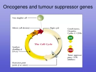
TUMOR-SUPPRESSOR GENES Molecular Oncology 2009. Michael Lea. TUMOR-SUPPRESSOR GENES - Lecture Outline. 1. Summary of tumor suppressor genes 2. P53 3. Rb 4. BRCA1 and 2 5. APC and DCC 6. PTEN and PPA2 7. LKB1 8. P16 9. WT1 and WTX 10. Epigenetic changes 11. miRNAs.

E N D
TUMOR-SUPPRESSOR GENESMolecular Oncology 2009 Michael Lea
TUMOR-SUPPRESSOR GENES - Lecture Outline 1. Summary of tumor suppressor genes 2. P53 3. Rb 4. BRCA1 and 2 5. APC and DCC 6. PTEN and PPA2 7. LKB1 8. P16 9. WT1 and WTX 10. Epigenetic changes 11. miRNAs
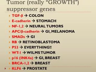
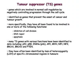
TUMOR-SUPPRESSOR GENES -Introduction Fusion of tumor cells with normal cells has been found to result in a loss of transformed properties. This suggests there are tumor suppressing activities in normal cells. Further support for this concept is provided by chromosomal deletions associated with some malignancies. The following is a list of tumor suppressor genes. Note there can be hereditary and sporadic defects for these genes. Gene Cancer type Hereditary syndrome APC Colon cancer Familial adenomatous polyposis BRCA1 Breast cancer BRCA2 Breast cancer DCC Colon cancer NF1 Neurofibromas Neurofibromatosis type 1 NF2 Schwannomas and Meningiomas Neurofibromatosis type 2 p53 Many types Li-Fraumeni syndrome PTEN Gliomas Rb Retinoblastoma Retinoblastoma VHL Kidney and other tumors von Hippel-Lindau syndrome WT1 Wilms tumor Wilms tumor In hereditary nonpolyposis colorectal cancer (HNPCC) defects have been noted in two genes coding for proteins used in DNA repair, namely MSH2 and MLH1. With defects in these genes there will be a high mutation frequency.
p53 Mutations in the p53 gene are found in a greater percentage of tumors than any other gene mutation. The situation with the p53 gene is complicated by the fact that mutation can result in 1. the loss of tumor suppressor function 2 oncogene activity including a dominant negative effect which overides the influence of the wild type gene. Hot spots have been identified in the p53 gene which are prone to mutation. Exposure to aflatoxin B1 causes a G->T transversion at codon 249 which is not generally seen in geographical regions with low exposure to aflatoxin. In the Li-Fraumeni syndrome, there is a germ-line mutation of the p53 gene resulting in a high incidence of cancer particularly tumors of the adrenal cortex, breast and brain and osteosarcomas. The p53 gene takes its name from the size of the 53 kd gene product. There are phosphorylation sites on the p53 protein including one which is phosphorylated by a cyclin-dependent kinase and which may be associated with cell cycle dependent translocation into the cell nucleus.
p53-activating signals and p53’s downstream effects
p53 The p53 protein is a transcriptional regulator that has been associated with blocking cell cycle progression and inducing apoptosis in some systems. These effects may be mediated by the products of genes whose expression is enhanced by the p53 protein including the p21WAF1/Cip1 gene and the Bax gene. The p21WAF1/Cip1 is known to be an inhibitor of cyclin-dependent kinase activity and can block cell cycle progression. The Bax protein is a promoter of apoptosis. The p53 gene is activated by DNA damage. It is thought to be important in normal cells to slow the cell cycle when DNA is damaged to permit DNA repair before the DNA is replicated. Failing this it may be preferable for the cell to die rather than perpetuate a damaged genome. Some of the action of the p53 gene on DNA repair may be mediated by activation of the Growth Arrest DNA damage gene, GADD45. the function of the p53 protein can be inhibited by binding to the product of the mdm-2 gene. This may constitute part of a feedback loop because the mdm-2 gene is activated by the p53 protein. When the mdm-2 gene is overexpressed as in some sarcomas it serves as an oncogene by supressing the function of the p53 protein.
Post-translational modification of p53 The p53 protein is subject to a variety of post-translational modifications. Phosphorylation and acetylation of p53 generally results in its stabilization and accumulation in the nucleus, followed by activation. Several protein kinases can phosphorylate p53. Mutant p53 is generally phosphorylated and acetylated at sites that are known to stabilize wild type p53 and could cause accumulation of dysfunctional p53 functioning as an oncogene. Overexpression of MDM2 E3 ubiquitin ligase results in the deactivation of p53 in many tumors. Reference: A.M. Bode and Z. Dong. Post-translational modification of p53 in tumorigenesis. Nature Reviews Cancer 4: 793-803, 2004.
Rb Retinoblastoma is an eye tumor of young children that occurs in a hereditary or a sporadic form. Deletions have been found in chromosome 13 associated with retinoblastoma. Inheritance of one defective gene puts the individual at greater risk. A somatic mutation in the other Rb gene will cause cancer whereas somatic mutations in two genes would be required in the normal individual. The Rb gene codes for a 105 kd protein. When hypophosphorylated p105 Rb exerts a growth restraining influence in the G1 phase of the cell cycle. Phosphorylation of the Rb protein inhibits its growth regulatory action. The Rb protein is a substrate for phosphorylation by cyclin-dependent kinases. Hyperphosphorylated Rb protein binds less tightly to the nucleus and less tightly to the E2F transcription factor which activates some genes for cell cycle progression. In the normal cell cycle, Rb becomes hyperphosphorylated at the G1/S transition and is released from the E2F transcription factor. The Rb protein can also bind specific DNA sequences and serve as a transcriptional regulator. Some transforming DNA viruses encode proteins that can bind with the Rb protein and block its function. These viral proteins include adenovirus E1A protein, SV40 large T antigen, E7 protein of human papilloma viruses 16 and 18 and polyoma middle T antigen. The Rb gene is required for normal development. Knockout mice die at about 14 to 15 days of embryonic development.
BRCA1 and BRCA2 GENES Mutations in the BRCA1 and BRCA2 genes impart increased susceptibility to breast cancer. Most cases are sporadic but some cases are familial. The BRCA1 gene codes for a large nuclear phophoprotein whose expression and phosphorylation is cell cycle dependent.It is probably a DNA-binding transcription factor and also involved in DNA repair. Mutations in the BRCA2 tumor-suppressor gene cause genomic instability and predisposition to cancer. BRCA2 appears to be required to prevent the breakdown of stalled replication forks. Disruption of this function leads to chromosomal rearrangements that occur spontaneously in dividing cells that have mutations in BRCA2. Reference: Lomonosov et al., Genes Dev. 17: 3017-3022 (2003)
APC In familial adenomatous polyposis (FAP) the colon is normal at birth, but during the first 20 years of life, hundreds of small polyps appear in the colon. The polyps are asymptomatic but there is a risk of progression to colon cancer that approaches 100% by age 50. The gene responsible is APC (adenomatous polyposis coli) which is involved in the degradation of beta-catenin.
PTEN (Phosphatase and Tensin homolog deleted on chromosome Ten) The PTEN gene is frequently mutated in human cancer, particularly gliomas. The PTEN protein can dephosphorylate phosphatidyl inositol 3,4,5 trisphosphate and thereby antagonize the phosphatidylinositol-3-kinase signaling pathway. PTEN negatively regulates intracellular levels of phosphatidylinositol-3,4,5-trisphosphate in cells and functions as a tumor suppressor by negatively regulating AKT/PKB signaling pathway. PTEN may also inhibit cell migration through protein phosphatase activity on a threonine phosphate residue. Reference: Raftopoulou et al. Science 303, 1179-1181 (2004).
PP2A PP2A is a serine/threonine phosphatase consisting in vivo of 3 subunits. The catalytic subunit (C-subunit) is present in 2 isoforms, a and b, which show the highest evolutionary conservation of all known enzymes, supporting the idea that they serve crucial functions. The catalytic subunit is constitutively associated with a structural/regulatory subunit (A-subunit), which exists in 2 isoforms encoded by different genes. The A-subunit is indispensable for the interaction of the catalytic subunit with the third regulatory subunit (B-subunit). Mutations in PPP2R1B, the gene encoding the beta-isoform of the A-subunit of PP2, have been recently described. The gene is localized on human chromosome 11q23, a region undergoing LOH in several tumors, including colon and lung cancer. Reference: Sloan-Kettering Institute > Cancer Biology & Genetics > Pier Paolo Pandolfi > Projects > Phosphatases and Cancer
PP2A Protein Phosphatase 2A (PP2A) plays a role in the critical cellular processes of protein synthesis, DNA replication, transcription, and metabolism. Small t antigen of SV40 interacts with the PP2A. This interaction reduces the ability of PP2A to inactivate ERK1 and MEK1 protein kinases, resulting in stimulation of proliferation of cells.
LKB1 Metformin and reduced risk of cancer in diabetic patients Metformin, widely given to patients with type 2 diabetes, works by targeting the enzyme AMPK (AMP activated protein kinase), which induces muscles to take up glucose from the blood. A recent breakthrough has found the upstream regulator of AMPK to be a protein kinase known as LKB1. LKB1 is a well recognized tumor suppressor. The Peutz-Jeghers tumor-suppressor gene encodes a protein-threonine kinase, LKB1, that phosphorylates and activates AMPK. Activation of AMPK by metformin and exercise requires LKB1, and this may also explain why exercise is beneficial in the primary and secondary prevention of certain cancers. Metformin use in patients with type 2 diabetes may reduce their risk of cancer. J.M.M. Evans, L.A. Donnelly, A.M. Emslie-Smith, D.R.Alessi and A.D. Morris. Brit. Med. J. 330:1304-1305 (2005), R.J. Shaw et al., Science 310: 1642-1646, 2005.
p16 The INK4a/ARF locus is of critical importance in tumor suppression. This locus is inactivated in about 40% of human cancers, a frequency only comparable with that of p53 inactivation. The INK4a/ARF locus encodes two tumor suppressors, p16INK4a and p14ARF/p19ARF (p14 when referred to the human protein and p19 when referred to the mouse protein), which share exons 2 and 3 but differ in their first exons and their respective promoters. Protein p16INK4a inhibits the activity of the CDK4,6/cycD kinases, thus contributing to the maintenance of the active, growth suppressive form of the retinoblastoma family of proteins. Matheu, A., Klatt, P., and Serrano, M. Regulation of the INK4a/ARF locus by histone deacetylase inhibitors. J. Biol. Chem. 280, 42433-42441, 2005
An X Chromosome Gene, WTX, is Commonly Inactivated in Wilm’s Tumor Wilm’s tumor is a pediatric kidney cancer associated with inactivation of the WT1 tumor suppressor gene in 5-10% of cases. The WTX gene is inactivated in approximately one third of Wilm’s tumors. In contrast to the biallelic inactivation of autosomal tumor-suppressor genes, WTX is inactivated by a monoallelic “single-hit” event targeting the single X chromosome in tumors in males and the active X chromosome in tumors from females. Reference: Rivera et al., Science 315: 642-645, 2007
Epigenetic Changes Some of the mechanisms involved in carcinogenesis may be epigenetic rather than genetic changes. Epigenetic changes include methylation of DNA and side-chain modification of histones including methylation and acetylation. One mechanism for the down regulation of tumor suppressor genes is methylation of promoter regions. Many groups are studying the combined action of inhibitors of DNA methylation and inhibitors of histone deacetylases as potential chemotherapeutic regimens.
miRNAs The deletion of the let-7 miRNA gene in C. elegans cause an uncontrolled proliferation of stem cells and overexpression of the ras gene. Lung cancer patients in Japan with the lowest levels of let-7 expression were found to have the worst prognosis. On the other hand, genes for some miRNAs may serve as oncogenes. A group of 13 miRNAs were reported to form a signature associated with prognosis and disease progression in patients with chronic lymphocytic leukemia (CLL). Reference: J. Couzin. A new cancer player takes the stage. Science 310: 766-767, 2005.
TUMOR-SUPPRESSOR GENES -Suggested reading B.H. Park and B. Vogelstein, In Holland-Frei Cancer Medicine 6th Edition, Part II, Section 1, 7. Tumor-Suppressor Genes, 2003. Robert Weinberg, The Biology of Cancer, Chapter 7, Garland Press, 2007.