Download
1 / 30
300 likes | 353 Vues
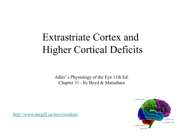
Extrastriate Cortex and Higher Cortical Deficits. Adler ’ s Physiology of the Eye 11th Ed. Chapter 31 - by Boyd & Matsubara. http://www.mcgill.ca/mvr/resident/. Multiple Visual Areas Beyond V1. Monkey Brain. Extrastriate Cortex. • Discrete cortical areas. • Hierarchical Organization

E N D
Extrastriate Cortex and Higher Cortical Deficits Adler’s Physiology of the Eye 11th Ed. Chapter 31 - by Boyd & Matsubara http://www.mcgill.ca/mvr/resident/
Multiple Visual Areas Beyond V1 Monkey Brain
Extrastriate Cortex • Discrete cortical areas • Hierarchical Organization -lower tier, higher tier • Parallel Streams - what vs. where - intra-area (blobs vs interblobs) - retinotopy • Feedforward and Feedback connections
Extrastriate Cortex Criteria For a Visual Area • Cyto-, myeloarchitecture • Connectivity New Way to Gain a Clear View of the Brain New York Times October 10, 2011 • Retinotopy -complete or partial map of visual space -represent a point in space only once? -smoothly varying? -orthogonal axes? • Specialized Function • Topography
Doctrine of the Receptive Field Retinotopy V2 V3 V1 Multiple “areas” V1, V2, V3
9 10 11 12 13 14 15 16 8 7 6 5 4 3 2 1 Receptive Fields at V1/V2 Border V2 V1 1 8 16
Functional Division of Labor? (motion) (color)
“What” versus “Where” Pathways Original concept came from lesion studies in monkeys (Mishkin & Ungerleider, 1982)
“What” versus “Where” Pathways Cross talk remains, and feedback is prevalent
Extrastriate Visual Areas thin- col - V4 inter- ori -V4 • V2 thick - ori dis - MT • V3d and V3A also magno-like • MT - MST magno input, V14B, thick, dir, dis, motion, depth • LIP - 7a Optic flow input, large RF, multimodal, project to frontal • V4 Central field input V1, V2, col ori, form primitives • IT input V2, V4, object features, face cells, project to multimodal Very large RF, object invariance, color constancy, training effects
MT * Strongly associated with motion perception Lesion and microstimulation studies in monkeys (Newsome and Pare, 1988; Salzman, Britten, Newsome, 1990)
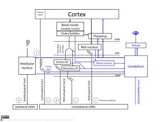
Wiring Diagram of Visual Areas “Subway Map From Hell” Van Essen et al., 1991
Human Visual Cortex MonkeyVisual Cortex Adlers, 2011
Human Lesion-Behavioral Correlations
Localization of Function in Humans Sources of information • Focal lesions • Histological Analysis • Hemispherectomy • Commissurotomy • Unilateral sodium amytal injection • Brain stimulation • Spontaneous and evoked electrical potentials • Functional brain imaging
Retinotopic Areas • Mapping Visual V1 with Clinical Stimulation (Dobelle et al, 1979) • Mapping Visual Areas Via Callosal Projections (Clark & Miklossy, 1990) • Mapping Visual V1 via Lesion-Scotoma Correlations (Horton & Hoyt, 1991) “state-of the-art” update of Gordon Holmes’Maps (1918) Human V1, V2, V3, V3A, V6, VP, V4, V8
Syndromes From Isolated Case Studies Akinetopsia Human MT Zihl et al., 1983
Syndromes From Isolated Case Studies Achromotopsia color constancy color constancy Human V4/V8
Visual Agnosia Aperceptive Agnosia - thought to be due to a disability in the construction of a stable representation of visual form, which impairs all high order recognition. Associative Agnosia - thought to reflect a deficit in accessing semantic (associative) knowledge about an object following the derivation of an intact perceptual representation of visual form. “Perception somehow striped of its meaning” (Teuber) Example: The man who mistook his wife for a hat (Oliver Sacks, 1985)
Syndromes From Isolated Case Studies Prosopagnosia
Syndromes From Isolated Case Studies Prosopagnosia Benton Facial Recognition Test
Human Face Areas anterior • fMRI studies with humans show increased activity in the fusiform face area (FFA). Inverted faces are hard to recognize. We are all face “experts” inflated brain inferior view
Artist’s rendition of spatial neglect German artist Anton Raderscheidt showed graduated recovery over eight-month period.
Modern Analysis of Lesion Overlap Right Hemisphere