Download
1 / 30
300 likes | 429 Vues
A comparison of membrane-containing bacteriophage and archaeal virus structures by cryo-EM. Sarah Butcher ERICE 2006. Enveloped virus session. irido. adeno. PRD1. Lineage 1 Lineage 2 Lineage 3. phi6. BTV. herpes. Membrane-containing viruses: antipasta misto.

E N D
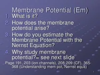
A comparison of membrane-containing bacteriophage and archaeal virus structures by cryo-EM Sarah Butcher ERICE 2006 Enveloped virus session
irido adeno PRD1 Lineage 1 Lineage 2 Lineage 3 phi6 BTV herpes
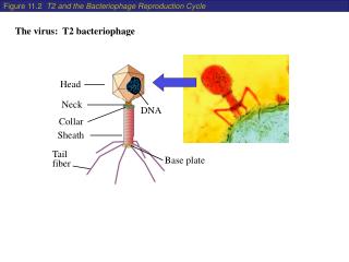
Membrane-containing viruses: antipasta misto Membrane required for host cell entry e.g. fusion with endosomes. Lipids derived from the host. Bacteriophage: Cystoviridae – f6 and f8 - fuse with Gram-negative host inner and outer membranes, releasing polymerase complex into cytoplasm Tectiviridae – PRD1 and Bam35 - fuse, releasing DNA + terminal proteins into cytoplasm Archaeal: SH1 - not known
Cryo-em studies • Relatively unstable viruses, so rapid preparation an advantage • Membrane structure preserved in native state • Possibility to look at mutants, protease treated, chemically disrupted • Resolution typically 8-12 Å, but varies within the reconstruction depending on the order. • Capsid proteins mainly peripheral membrane proteins • Integral membrane proteins occasionally revealed • Image reconstruction generally relies on icosahedral symmetry for orientation determination and averaging. Hence symmetry mismatches will not be resolved.
f6 entry Pseudomonas syringae CM OM PG
3D reconstruction of f6 and f8 virion Jäälinoja et al. (in preparation)
Structure of the f6 nucleocapsid • Three-dimensional reconstruction from cryo-EM dataapplying icosahedralsymmetry • Two protein shells • Hexameric packagingmotor at the vertices(symmetry-mismatch)
25 Å 14 Å PRD1 cryo-EM 12 Å Butcher et al. 1995 EMBO J. San Martin et al. 2001 Structure San Martin et al. 2002 NSB
Major capsid protein is a peripheral membrane protein R=36% R=34% R=31% Benson et al, 1999 Cell; San Martin et al. 2001 Structure
EM/Xray combination Benson et al, 1999 Cell; San Martin et al. 2001 Structure; San Martin et al. 2002 NSB
Bam35 • Infects Gram-positive host Bacillus thuringiensis • Very limited sequence similarity to bacteriophage PRD1 • Address structural similarity using cryo-EM and X-ray data from PRD1
7.3 Å icosahedral reconstruction of bacteriophage Bam35 • Reconstructions calculated from both full virion and particle lacking DNA Laurinmäki et al. (2005) Structure
Modelling with homologous proteins Laurinmäki et al. (2005) Structure
Bam35 membrane proteins Laurinmäki et al. (2005) Structure
Archaeal virus SH1 800Å 100Å Jäälinoja et al. (in preparation)
Density distribution and resolution Capsid DNA Spike Membrane Jäälinoja et al. (in preparation)
Archaeal virus SH1 Jäälinoja et al. (in preparation)
Unusual capsomers • 2- and 3–fold symmetric capsomers on capsid surface. • Quasi-hexagonal capsomer base. • Slightly skewed at 2f and adjacent capsomers, not elsewhere. • All with similar mass Jäälinoja et al. (in preparation)
Structure at 5-fold axis • Very weak signal from peripheral domain. • Possible symmetry mismatch in external domains. • Transmembrane complex. Jäälinoja et al. (in preparation)
Vertex Reconstruction Orientation angles and vertex positions derived from output of an icosahedral reconstruction. Vertices are compared and classified revealing differences in composition and orientation. Subsets of vertices can be used for reconstruction and any desired symmetry can be applied.
SH1 spike reconstruction Briggs et al. (2005) JSB; Huiskonen et al. (submitted); Jäälinoja et al. (in preparation)
Cryo-em studies – conclusions • Capsid proteins mainly peripheral membrane proteins, connected by a-helices • Integral membrane proteins occasionally revealed, so far apparently a-helical • Membranes are icosahedral, locally affected by membrane proteins
Acknowledgements • Benita Koli • Dennis Bamford • Hanna Kivelä • John Briggs • Stephen Fuller